
To label:
To label the diagram of the antibody.
Introduction:
The structure of the antibody resembles the alphabet Y and consists of 4 proteins (2 light chains and 2 heavy chains).
Pictorial representation:
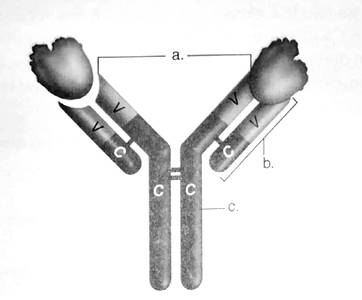
Figure: The structure of an antibody molecule.
The correct answers are:
- Antigen binding sites
- Light chain
- Heavy chain
V stands for the variable region
C stands for the constant region
Explanation for the answers:
- Antigen binding site − These are the sites on the amino-terminal end of both light and heavy chain. It provides a site of attachment for the antigen.
- Light chain − There is 2 identical light chains consisting of polypeptides. They are bound to the heavy chain via disulfide bonds and non-covalent interactions.
- Heavy chain − There are 5 types of heavy chains (accordingly antibodies are named) and attached to each other via the disulfide bonds.
V stands for variable region. The amino-terminal end of both heavy and light chain of about 100-110 amino acids vary considerably among antibodies and hence are called as variable region.
C stands for the constant region. The carboxy-terminalend of both light and heavy chain shows similarity among antibodies and hence is considered as constant regions.
Want to see the full answer?
Check out a sample textbook solution
Chapter 13 Solutions
CUSTOMIZED LAB MANUAL FOR BIOLOGY 1406

- When setting up a PCR reaction to act as a negative control for the surface protein A gene... Which primers will you add to the reaction mix? mecA primers, spa primers, mecA primers and spa primers, no primers What will you add in place of template? sterile water, MRSA DNA, Patient DNA, S. aureus DNAarrow_forwardDraft a science fair project for a 11 year old based on the human body, specifically the liverarrow_forwardYou generate a transgenic mouse line with a lox-stop-lox sequence upstream of a dominant-negative Notch fused to GFP. Upon crossing this mouse with another mouse line expressing ectoderm-specific Cre, what would you expect for the phenotype of neuronal differentiation in the resulting embryos?arrow_forward
- Hair follicle formation is thought to result from a reaction-diffusion mechanism with Wnt and its antagonist Dkk1. How is Dkk1 regulated by Wnt? Describe specific cis-regulatory elements and the net effect on Dkk1 expression.arrow_forwardLimetown S1E4 Transcript: E n 2025SP-BIO-111-PSNT1: Natu X Natural Selection in insects X + newconnect.mheducation.com/student/todo CA NATURAL SELECTION NATURAL SELECTION IN INSECTS (HARDY-WEINBERG LAW) INTRODUCTION LABORATORY SIMULATION A Lab Data Is this the correct allele frequency? Is this the correct genotype frequency? Is this the correct phenotype frequency? Total 1000 Phenotype Frequency Typica Carbonaria Allele Frequency 9 P 635 823 968 1118 1435 Color Initial Frequency Light 0.25 Dark 0.75 Frequency Gs 0.02 Allele Initial Allele Frequency Gs Allele Frequency d 0.50 0 D 0.50 0 Genotype Frequency Moths Genotype Color Moths Released Initial Frequency Frequency G5 Number of Moths Gs NC - Xarrow_forwardWhich of the following is not a sequence-specific DNA binding protein? 1. the catabolite-activated protein 2. the trp repressor protein 3. the flowering locus C protein 4. the flowering locus D protein 5. GAL4 6. all of the above are sequence-specific DNA binding proteinsarrow_forward
- Which of the following is not a DNA binding protein? 1. the lac repressor protein 2. the catabolite activated protein 3. the trp repressor protein 4. the flowering locus C protein 5. the flowering locus D protein 6. GAL4 7. all of the above are DNA binding proteinsarrow_forwardWhat symbolic and cultural behaviors are evident in the archaeological record and associated with Neandertals and anatomically modern humans in Europe beginning around 35,000 yBP (during the Upper Paleolithic)?arrow_forwardDescribe three cranial and postcranial features of Neanderthals skeletons that are likely adaptation to the cold climates of Upper Pleistocene Europe and explain how they are adaptations to a cold climate.arrow_forward
- Biology Questionarrow_forward✓ Details Draw a protein that is embedded in a membrane (a transmembrane protein), label the lipid bilayer and the protein. Identify the areas of the lipid bilayer that are hydrophobic and hydrophilic. Draw a membrane with two transporters: a proton pump transporter that uses ATP to generate a proton gradient, and a second transporter that moves glucose by secondary active transport (cartoon-like is ok). It will be important to show protons moving in the correct direction, and that the transporter that is powered by secondary active transport is logically related to the proton pump.arrow_forwarddrawing chemical structure of ATP. please draw in and label whats asked. Thank you.arrow_forward
 Human Anatomy & Physiology (11th Edition)BiologyISBN:9780134580999Author:Elaine N. Marieb, Katja N. HoehnPublisher:PEARSON
 Biology 2eBiologyISBN:9781947172517Author:Matthew Douglas, Jung Choi, Mary Ann ClarkPublisher:OpenStax
 Anatomy & PhysiologyBiologyISBN:9781259398629Author:McKinley, Michael P., O'loughlin, Valerie Dean, Bidle, Theresa StouterPublisher:Mcgraw Hill Education,
 Molecular Biology of the Cell (Sixth Edition)BiologyISBN:9780815344322Author:Bruce Alberts, Alexander D. Johnson, Julian Lewis, David Morgan, Martin Raff, Keith Roberts, Peter WalterPublisher:W. W. Norton & Company
 Laboratory Manual For Human Anatomy & PhysiologyBiologyISBN:9781260159363Author:Martin, Terry R., Prentice-craver, CynthiaPublisher:McGraw-Hill Publishing Co.
 Inquiry Into Life (16th Edition)BiologyISBN:9781260231700Author:Sylvia S. Mader, Michael WindelspechtPublisher:McGraw Hill Education