Human Anatomy & Physiology (11th Edition)
11th Edition
ISBN:9780134580999
Author:Elaine N. Marieb, Katja N. Hoehn
Publisher:Elaine N. Marieb, Katja N. Hoehn
Chapter1: The Human Body: An Orientation
Section: Chapter Questions
Problem 1RQ: The correct sequence of levels forming the structural hierarchy is A. (a) organ, organ system,...
Related questions
Question
Refer to the figures above:
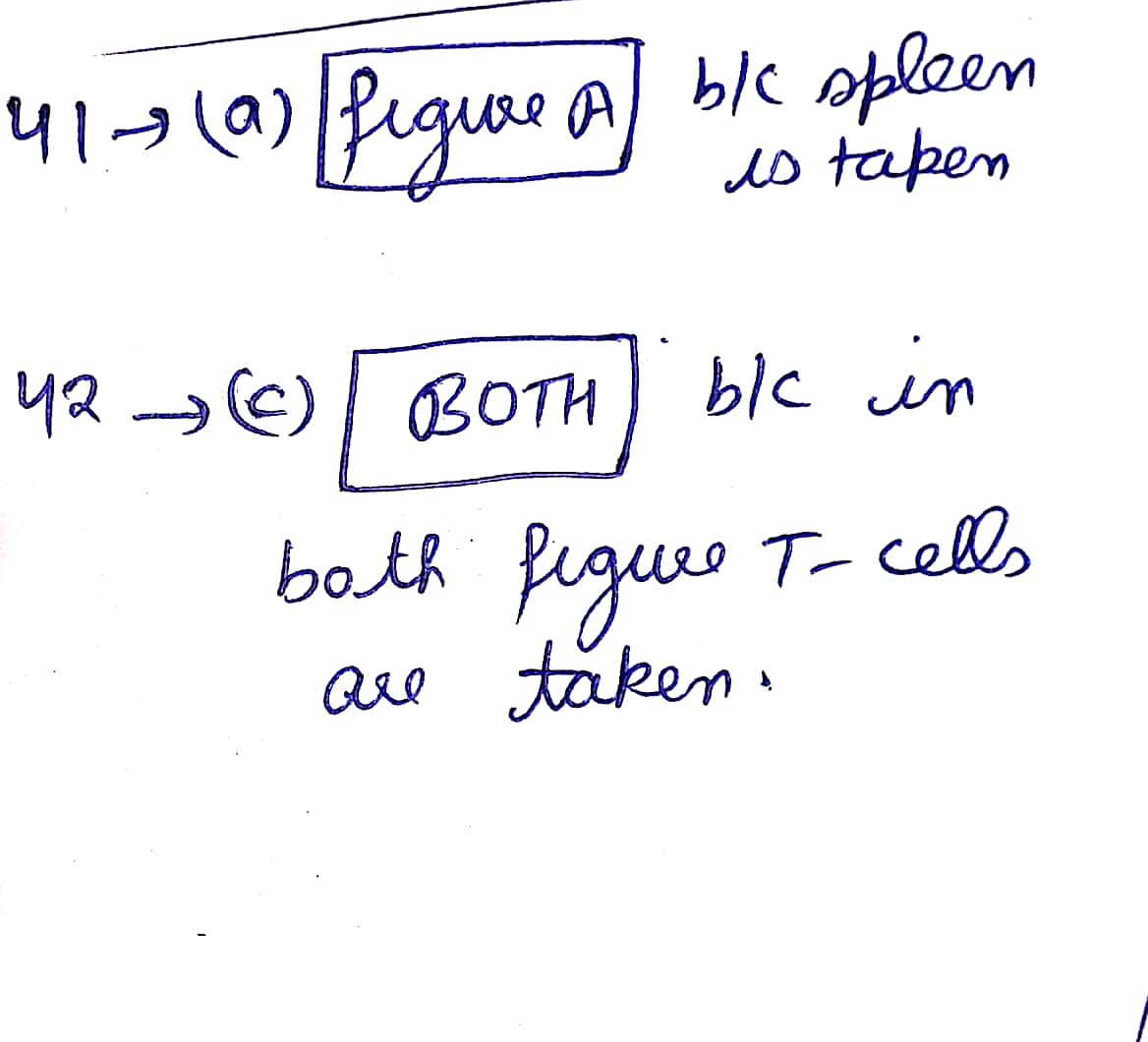
41) Which figure displays the rate of bacterial growth in a living animal?
a) Figure A
b) Figure C
C) Both
d) Neither
e) Cannot be determined
42) Which figure tests the effects of one or more lymphocytes?
a) Figure A
b) Figure C
c) Both
d) Neither
e) Cannot be determined
43) Which figure demonstrates that addition of effector PCs leads to bacterial death?
a) Figure A
b) Figure C
C) Both
d) Neither
e) Cannot be determined
44) Based on these figures, Listeria infection control is most directly due to which effector cell:
a) CTL
b) T helper
c) Macrophage
d) Basophil
d) None of the above

Transcribed Image Text:### Transcription for Educational Purposes
#### Figure A: T Lymphocytes Adoptively Transfer Specific Immunity
- **Y-axis**: Number of viable *Listeria* organisms in spleen (log10)
- **X-axis**: Days post-infection
- **Key**:
- Brown line with crosses: Immune T cells
- Blue line with squares: Nonimmune T cells
This graph shows that the number of *Listeria* organisms in the spleen increases over time when nonimmune T cells are present, while it remains low with immune T cells.
#### Figure C: Only Activated Macrophages Kill *Listeria* In Vitro
- **Y-axis**: Percentage killing of *Listeria* in vitro
- **X-axis**: Leukocytes added (×10^6)
- **Key**:
- Purple line with crosses: Immune T cells
- Orange line with squares: Resting macrophages
- Green line with triangles: Activated macrophages
This graph illustrates how different types of immune cells kill *Listeria* in vitro. Activated macrophages show a significant increase in killing efficiency as the number of leukocytes increases.
#### Questions
41) Which figure displays the rate of bacterial growth in a living animal?
- a) Figure A
- b) Figure C
- c) Both
- d) Neither
- e) Cannot be determined
42) Which figure tests the effects of one or more lymphocytes?
- a) Figure A
- b) Figure C
- c) Both
- d) Neither
- e) Cannot be determined
43) Which figure demonstrates that addition of effector APCs leads to bacterial death?
- a) Figure A
- b) Figure C
- c) Both
- d) Neither
- e) Cannot be determined
44) Based on these figures, *Listeria* infection control is most directly due to which effector cell:
- a) CTL
- b) T helper
- c) Macrophage
- d) Basophil
- e) None of the above
These graphs and accompanying questions are designed to help students understand the role of specific immune cells in controlling *Listeria* infections.
Expert Solution
Step 1

Step by step
Solved in 2 steps with 2 images

Knowledge Booster
Learn more about
Need a deep-dive on the concept behind this application? Look no further. Learn more about this topic, biology and related others by exploring similar questions and additional content below.Recommended textbooks for you

Human Anatomy & Physiology (11th Edition)
Biology
ISBN:
9780134580999
Author:
Elaine N. Marieb, Katja N. Hoehn
Publisher:
PEARSON

Biology 2e
Biology
ISBN:
9781947172517
Author:
Matthew Douglas, Jung Choi, Mary Ann Clark
Publisher:
OpenStax

Anatomy & Physiology
Biology
ISBN:
9781259398629
Author:
McKinley, Michael P., O'loughlin, Valerie Dean, Bidle, Theresa Stouter
Publisher:
Mcgraw Hill Education,

Human Anatomy & Physiology (11th Edition)
Biology
ISBN:
9780134580999
Author:
Elaine N. Marieb, Katja N. Hoehn
Publisher:
PEARSON

Biology 2e
Biology
ISBN:
9781947172517
Author:
Matthew Douglas, Jung Choi, Mary Ann Clark
Publisher:
OpenStax

Anatomy & Physiology
Biology
ISBN:
9781259398629
Author:
McKinley, Michael P., O'loughlin, Valerie Dean, Bidle, Theresa Stouter
Publisher:
Mcgraw Hill Education,

Molecular Biology of the Cell (Sixth Edition)
Biology
ISBN:
9780815344322
Author:
Bruce Alberts, Alexander D. Johnson, Julian Lewis, David Morgan, Martin Raff, Keith Roberts, Peter Walter
Publisher:
W. W. Norton & Company

Laboratory Manual For Human Anatomy & Physiology
Biology
ISBN:
9781260159363
Author:
Martin, Terry R., Prentice-craver, Cynthia
Publisher:
McGraw-Hill Publishing Co.

Inquiry Into Life (16th Edition)
Biology
ISBN:
9781260231700
Author:
Sylvia S. Mader, Michael Windelspecht
Publisher:
McGraw Hill Education