
Videos
Describe the actions of the frog heart chambers during a cardiac cycle.
To write:
The actions of the heart chambers during a cardiac cycle in frog.
Introduction:
Heart is the vital organ of the circulatory system as it pumps blood all over the body. Human heart consists of four chambers: two upper chambers or atria and two lower chambers or ventricles. Blood passes across a valve before parting each heart chamber.
Explanation of Solution
Heart of frog:
The heart in frog consists of three chambers. There are two atrial chambers which drain into a single and common ventricle.
The pacemaker in the frog’s heart is present in the posterior side of the heart. The pacemaker is known as the sinus venosus. This sinus venosus is similar to the sinoatrial node inside the cardiac tissue of human.
Actions of frog heart chambers during a cardiac cycle are:
There occurs an atrial systole or atrial contraction. Then there is an atrial diastole or relaxation. This diastole is followed by a ventricle systole. Then ventricular systole is followed by a ventricle diastole. These actions complete one heart beat in the frog.
Blood circulation in frog by the action of the heart chambers is:
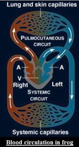
The three chambers in the frog’s heart are two atria and one ventricle. Blood exits the heart by the ventricle across a single truncus arteriosus. It is a short blood vessel that immediately bifurcates into two aortic arches (left and right) dorsal to the heart. These aortic arches then reunite and form a single aorta in the mid-dorsal part of the body cavity. Each aortic arch gives off a branch that runs to the lungs and skin where the oxygenation of blood takes place. Carotid arteries also divide into the aortic arches. These deliver to the head region.
Veins transport blood back to the left and right atria. Both the atria then drain into a single ventricle. Then again the blood from the ventricle enters into the pulmocutaneous or body circulation. As, there exists a single ventricle, there occurs some mixing of the oxygenated and deoxygenated blood. Ventricular folds are present that prevent a complete combination of the venous and arterial blood.
So, actions of the chambers of the frog heart involve an atrial systole followed by an atrial diastole. Atrial diastole is then followed by a ventricle systole which is further followed by a ventricle diastole.
Hence, actions of the chambers of the frog heart during a cardiac cycle involve an atrial systole followed by an atrial diastole. Atrial diastole is then followed by a ventricle systole which is further followed by a ventricle diastole.
Want to see more full solutions like this?
Chapter S3 Solutions
LAB MANUAL FOR HUMAN A&P MAIN VERSION


- Pleiotropic genes are genes that (blank) Cause a swapping of organs/structures, are the result of duplicated sets of chromosomes, never produce protein products, and have more than one purpose/functionarrow_forwardA loss of function mutation in Pitx1 enhancers can cause (blank) Removal of Pitx1 exons and growth of ectopic hindlimbs, growth of extra ectopic forelimbs, loss of forelimb specification and development, and loss of hindlimb specification and developmentarrow_forwardHox1a most likely contributes to (blank) patterning in the developing embryo? Ventral, posterior, limb or anteriorarrow_forward
- Select all of the following that can help establish Hox gene expression boundaries (things that affect Hox and not things that Hox affects). Retinoic acid, anterior/posterior axis, fibroblast growth factors, vagal neural crest, and enhancersarrow_forwardEctopic expression of Hox often results in (blank) phenotypes. (Blank) transformations are characterized by the replacement of one body part/structure with another. Hoxeotic, homealoneotic, joexotic, or homeoticarrow_forwardWhat's the difference when drawing omega-6 and omega-3?arrow_forward
- . Consider a base substitution mutation that occurred in a DNA sequence that resulted in a change in the encoded protein from the amino acid glutamic acid to aspartic acid. Normally the glutamic acid amino acid is located on the outside of the soluble protein but not near an active site. O-H¨ A. What type of mutation occurred? O-H B. What 2 types of chemical bonds are found in the R-groups of each amino acid? The R groups are shaded. CH2 CH2 CH2 H2N-C-COOH H2N-C-COOH 1 H Glutamic acid H Aspartic acid C. What 2 types of bonds could each R-group of each of these amino acids form with other molecules? D. Consider the chemical properties of the two amino acids and the location of the amino acid in the protein. Explain what effect this mutation will have on this protein's function and why.arrow_forwardengineered constructs that consist of hollow fibers are acting as synthetic capillaries, around which cells have been loaded. The cellular space around a single fiber can be modeled as if it were a Krogh tissue cylinder. Each fiber has an outside “capillary” radius of 100 µm and the “tissue” radius can be taken as 200 µm. The following values apply to the device:R0 = 20 µM/secaO2 = 1.35 µM/mmHgDO2,T = 1.67 x 10⁻⁵ cm2/secPO2,m = 4 x 10⁻³ cm/secInstead of blood inside the fibers, the oxygen transport and tissue consumption are being investigated by usingan aqueous solution saturated with pure oxygen. As a result, there is no mass transfer resistance in the synthetic“capillary”, only that due to the membrane itself. Rather than accounting for pO2 variations along the length ofthe fiber, use an average value in the “capillary” of 130 mmHg.Is the tissue fully oxygenated?arrow_forwardMolecular Biology Please help with question. thank you You are studying the expression of the lac operon. You have isolated mutants as described below. In the presence of glucose, explain/describe what would happen, for each mutant, to the expression of the lac operon when you add lactose AND what would happen when the bacteria has used up all of the lactose (if the mutant is able to use lactose).5. Mutations in the lac operator that strengthen the binding of the lac repressor 200 fold 6. Mutations in the promoter that prevent binding of RNA polymerase 7. Mutations in CRP/CAP protein that prevent binding of cAMP8. Mutations in sigma factor that prevent binding of sigma to core RNA polymerasearrow_forward
- Molecular Biology Please help and there is an attached image. Thank you. A bacteria has a gene whose protein/enzyme product is involved with the synthesis of a lipid necessary for the synthesis of the cell membrane. Expression of this gene requires the binding of a protein (called ACT) to a control sequence (called INC) next to the promoter. A. Is the expression/regulation of this gene an example of induction or repression?Please explain:B. Is this expression/regulation an example of positive or negative control?C. When the lipid is supplied in the media, the expression of the enzyme is turned off.Describe one likely mechanism for how this “turn off” is accomplished.arrow_forwardMolecular Biology Please help. Thank you. Discuss/define the following:(a) poly A polymerase (b) trans-splicing (c) operonarrow_forwardMolecular Biology Please help with question. Thank you in advance. Discuss, compare and contrast the structure of promoters inprokaryotes and eukaryotes.arrow_forward
 Concepts of BiologyBiologyISBN:9781938168116Author:Samantha Fowler, Rebecca Roush, James WisePublisher:OpenStax College
 Comprehensive Medical Assisting: Administrative a...NursingISBN:9781305964792Author:Wilburta Q. Lindh, Carol D. Tamparo, Barbara M. Dahl, Julie Morris, Cindy CorreaPublisher:Cengage Learning
 Human Biology (MindTap Course List)BiologyISBN:9781305112100Author:Cecie Starr, Beverly McMillanPublisher:Cengage Learning
 Human Physiology: From Cells to Systems (MindTap ...BiologyISBN:9781285866932Author:Lauralee SherwoodPublisher:Cengage Learning
 Biology: The Dynamic Science (MindTap Course List)BiologyISBN:9781305389892Author:Peter J. Russell, Paul E. Hertz, Beverly McMillanPublisher:Cengage Learning