Human Biology Custom Edition
11th Edition
ISBN: 9781337631532
Author: Cecie Starr, Beverly McMillan
Publisher: Cengage Learning
expand_more
expand_more
format_list_bulleted
Concept explainers
Textbook Question
Chapter 7, Problem 7RQ
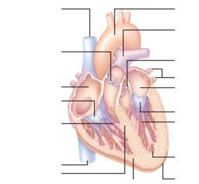
Label the heart’s main parts in the diagram below.

Expert Solution & Answer
Trending nowThis is a popular solution!

Students have asked these similar questions
4. This question focuses on entrainment.
a. What is entrainment?
b. What environmental cues are involved in entrainment, and which one is most influential?
c. Why is entrainment necessary?
d. Assuming that a flash of darkness is an effective zeitgeber, what impact on circadian
rhythms would you expect to result from an event such as the 2024 solar eclipse (assume
it was viewed from Carbondale IL, where totality occurred at about 2 pm)? Explain your
reasoning. You may wish to consult this phase response diagram.
Phase Shift (Hours)
Delay Zone
Advance Zone
Dawn
Mid-day
Dusk
Night
Dawn
Time of Light Stimulus
e. Finally, give a real-world example of how knowledge of circadian rhythms and
entrainment has implications for human health and wellbeing or conservation biology.
This example could be from your reading or from things discussed in class.
Generate one question that requires a Punnet Squre to solve the question. Then show how you calculate the possibilities of genotype and phenotype
Briefly state the physical meaning of the electrocapillary equation (Lippman equation).
Chapter 7 Solutions
Human Biology Custom Edition
Ch. 7 - List the functions of the cardiovascular system.Ch. 7 - Define a heartbeat, giving the sequence of events...Ch. 7 - What is the difference between the systemic and...Ch. 7 - Prob. 4RQCh. 7 - Prob. 5RQCh. 7 - State the main functions of venules and veins....Ch. 7 - Label the hearts main parts in the diagram below.Ch. 7 - Cells obtain nutrients from and deposit waste into...Ch. 7 - The contraction phase of the heartbeat is ______;...Ch. 7 - Prob. 3SQ
Ch. 7 - In the systemic circuit, the hearts _______ half...Ch. 7 - After you eat, blood passing through the GI tract...Ch. 7 - Prob. 6SQCh. 7 - Prob. 7SQCh. 7 - Prob. 8SQCh. 7 - _____ contraction drives blood through the...Ch. 7 - Match the type of blood vessel with its major...Ch. 7 - Prob. 11SQCh. 7 - A patient suffering from hypertension may receive...Ch. 7 - Heavy smokers often develop abnormally high blood...Ch. 7 - Prob. 3CTCh. 7 - Several years ago the deaths of several airline...
Knowledge Booster
Learn more about
Need a deep-dive on the concept behind this application? Look no further. Learn more about this topic, biology and related others by exploring similar questions and additional content below.Similar questions
- Explain in a small summary how: What genetic information can be obtained from a Punnet square? What genetic information cannot be determined from a Punnet square? Why might a Punnet Square be beneficial to understanding genetics/inheritance?arrow_forwardIn a small summary write down:arrow_forwardNot part of a graded assignment, from a past midtermarrow_forward
- Noggin mutation: The mouse, one of the phenotypic consequences of Noggin mutationis mispatterning of the spinal cord, in the posterior region of the mouse embryo, suchthat in the hindlimb region the more ventral fates are lost, and the dorsal Pax3 domain isexpanded. (this experiment is not in the lectures).a. Hypothesis for why: What would be your hypothesis for why the ventral fatesare lost and dorsal fates expanded? Include in your answer the words notochord,BMP, SHH and either (or both of) surface ectoderm or lateral plate mesodermarrow_forwardNot part of a graded assignment, from a past midtermarrow_forwardNot part of a graded assignment, from a past midtermarrow_forward
arrow_back_ios
SEE MORE QUESTIONS
arrow_forward_ios
Recommended textbooks for you
 Human Biology (MindTap Course List)BiologyISBN:9781305112100Author:Cecie Starr, Beverly McMillanPublisher:Cengage Learning
 Human Physiology: From Cells to Systems (MindTap ...BiologyISBN:9781285866932Author:Lauralee SherwoodPublisher:Cengage Learning
 Biology 2eBiologyISBN:9781947172517Author:Matthew Douglas, Jung Choi, Mary Ann ClarkPublisher:OpenStax
 Fundamentals of Sectional Anatomy: An Imaging App...BiologyISBN:9781133960867Author:Denise L. LazoPublisher:Cengage Learning

Human Biology (MindTap Course List)
Biology
ISBN:9781305112100
Author:Cecie Starr, Beverly McMillan
Publisher:Cengage Learning

Human Physiology: From Cells to Systems (MindTap ...
Biology
ISBN:9781285866932
Author:Lauralee Sherwood
Publisher:Cengage Learning


Biology 2e
Biology
ISBN:9781947172517
Author:Matthew Douglas, Jung Choi, Mary Ann Clark
Publisher:OpenStax

Fundamentals of Sectional Anatomy: An Imaging App...
Biology
ISBN:9781133960867
Author:Denise L. Lazo
Publisher:Cengage Learning

Dissection Basics | Types and Tools; Author: BlueLink: University of Michigan Anatomy;https://www.youtube.com/watch?v=-_B17pTmzto;License: Standard youtube license