
Concept explainers
(a).
To label: The photomicrographs in figure 6.3.
Introduction: The epithelial tissue is formed by joining the epithelial cells in the body. The epithelial cells have important functions like protecting the tissue layers and hence they are always found on the surface of the organs and tissues. The epithelial cells also act as a barrier to control the movement of molecules in-between the glands.
(a).
Answer to Problem 1.3BGL
Pictorial representation:
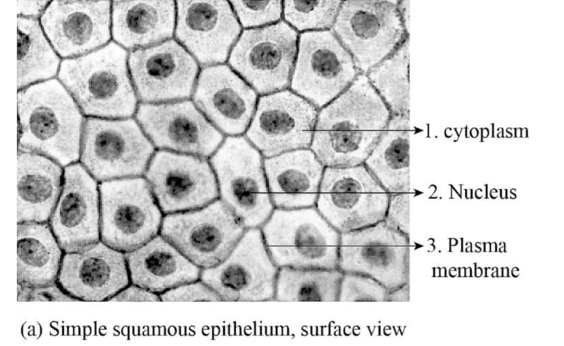
Fig.1: The figure showing simple squamous epithelial cell.
Explanation of Solution
Simple squamous epithelial cells: The simple squamous epithelial cells are a single layer of epithelial cells that are found in the alveoli in the lungs, glomerular capsule; as endothelium in the inner lining of capillaries, as mesothelium in the peritoneal cavity and mediastinum. These epithelial cells have a specific function of gaseous exchange by diffusion, secretion, filtration, absorption. The presence of single layer allows easy passage of the gases in-between the cells.
(b).
To label: The photomicrographs in figure 6.4.
(b).
Answer to Problem 1.3BGL
Pictorial representation:
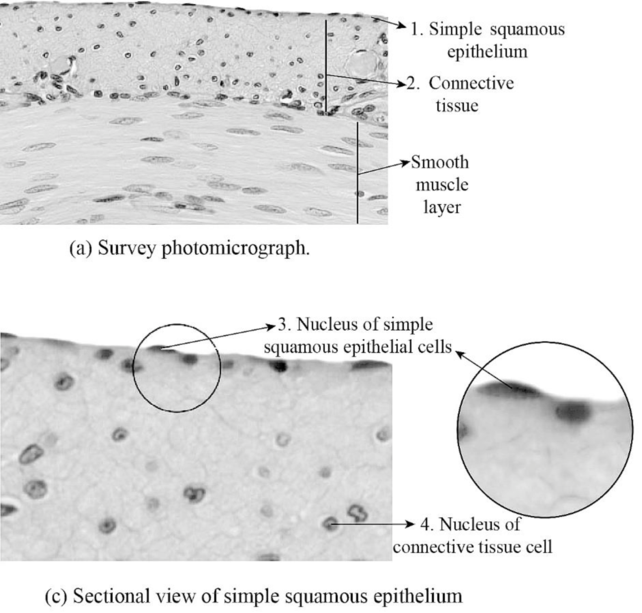
Fig.2: The figure showing sectional view of simple squamous epithelial cell.
Explanation of Solution
The cross section image of the simple squamous epithelial cells shows, smooth muscle are covered by the epithelial cells. The connective tissue forms the basement membrane for the epithelial cells to form a single layer of simple squamous epithelial cell over the smooth muscle.
(c).
To label: The photomicrographs in figure 6.5.
(c).
Answer to Problem 1.3BGL
Pictorial representation:
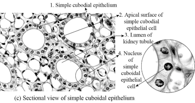
Fig.3: The figure showing simple cuboidal epithelial cell.
Explanation of Solution
Simple cuboidal epithelial cells: The simple cuboidal epithelial cells are a single layer of epithelial cells that are found in the wall of kidney tubules and glands. These epithelial cells have a specific function of absorption and secretion. These epithelial cells absorb substance from outside of the tubules and secret those substances inside the kidney tubules.
(d).
To label: The photomicrographs in figure 6.6.
(d).
Answer to Problem 1.3BGL
Pictorial representation:
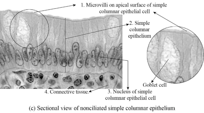
Fig.4: The figure showing simple columnar epithelial cell.
Explanation of Solution
Simple columnar epithelial cells: The simple columnar epithelial cells are a single layer of epithelial cells that are found in the lining of the stomach and intestines. These are special types of cells that are elongated and have microvilli present on them. The columnar epithelial cells have special functions like secreting digestive juices and absorption by increasing the surface area in the small intestine.
(e).
To label: The photomicrographs in figure 6.7.
(e).
Answer to Problem 1.3BGL
Pictorial representation:
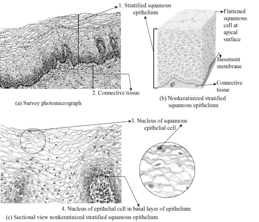
Fig.5: The figure showing stratified squamous epithelial cell.
Explanation of Solution
Stratified squamous epithelium: The stratified squamous epithelium cells are found at areas of lips, esophagus, mouth, vagina, and anus. The stratified epithelium formed by multiple layers of epithelium cells that makes surface area to be thicker, damage resistance, forms a protective barrier against friction and abrasion, and also prevent the entry of pathogens due to their tight adherence of multi-layered cells.
(f).
To label: The photomicrographs in figure 6.8.
(f).
Answer to Problem 1.3BGL
Pictorial representation:
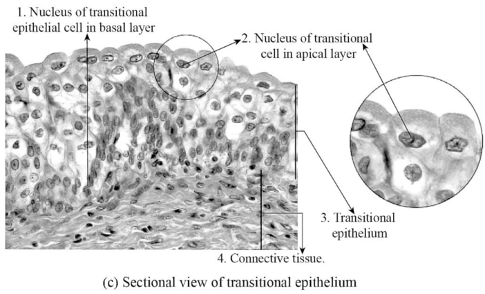
Fig.6: The figure showing transitional epithelial cell.
Explanation of Solution
Transitional epithelium cell: The transitional epithelial cells are one of the types of epithelial cells that has a special function like changing the structure of the cell. The transitional epithelial cells are found in the lining of the urinary bladder, urethra, and ureters. The function of the transitional epithelial cells is to form a protective barrier on the surface of the urinary bladder and also reduces tension.
(g).
To label: The photomicrographs in figure 6.9.
Introduction: The epithelial cell is formed by joining cells that together form layers of epithelial cells in the body. The epithelial cells have important functions like protecting the tissue layers and hence they are always found on the surface of the organs and tissues.
(g).
Answer to Problem 1.3BGL
Pictorial representation:
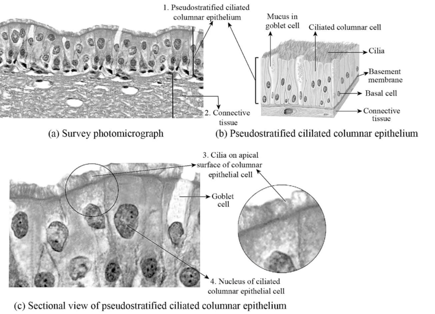
Fig.7: The figure showing pseudostratified epithelial cell.
Explanation of Solution
Pseudostratified epithelium cell: The stratified epithelium formed by multiple layers of epithelium cells that makes surface area to be thicker, damage resistance, forms a protective barrier against friction and abrasion, and also prevent the entry of pathogens. The pseudostratified epithelial cells also look like multilayer epithelium but it is a single-layered thick cell. The pseudostratified columnar epithelial cells found in the nasal cavity lining. The pseudostratified columnar epithelial cells protect the nasal cavity by secretion of mucus to trap the pathogens and dust.
Want to see more full solutions like this?
Chapter 6 Solutions
Laboratory Manual for Anatomy and Physiology, 6e Loose-Leaf Print Companion with WileyPLUS Blackboard Card Set


- Please indentify the unknown organismarrow_forwardPlease indentify the unknown organismarrow_forward5G JA ATTC 3 3 CTIA A1G5 5 GAAT I I3 3 CTIA AA5 Fig. 5-3: The Eco restriction site (left) would be cleaved at the locations indicated by the arrows. However, a SNP in the position shown in gray (right) would prevent cleavage at this site by EcoRI One of the SNPs in B. rapa is found within the Park14 locus and can be detected by RFLP analysis. The CT polymorphism is found in the intron of the Bra013780 gene found on Chromosome 1. The Park14 allele with the "C" in the SNP has two EcoRI sites and thus is cleaved twice by EcoRI If there is a "T" in that SNP, one of the EcoRI sites is altered, so the Park14 allele with the T in the SNP has only one EcoRI site (Fig. 5-3). Park14 allele with SNP(C) Park14 allele with SNPT) 839 EcoRI 1101 EcoRI 839 EcoRI Fig. 5.4: Schematic restriction maps of the two different Park14 alleles (1316 bp long) of B. rapa. Where on these maps is the CT SNP located? 90 The primers used to amplify the DNA at the Park14 locus (see Fig. 5 and Table 3 of Slankster et…arrow_forward
- From your previous experiment, you found that this enhancer activates stripe 2 of eve expression. When you sequence this enhancer you find several binding sites for the gap gene, Giant. To test how Giant interacts with eve, you decide to remove all of the Giant binding sites from the eve enhancer. What results do you expect to see with respect to eve expression?arrow_forwardWhat experiment could you do to see if the maternal gene, bicoid, is sufficient to form anterior fates?arrow_forwardYou’re curious about the effect that gap genes have on the pair-rule gene, evenskipped (eve), so you isolate and sequence each of the eve enhancers. You’re particularly interested in one of the enhancers, which is just upstream of the eve gene. Describe an experimental technique you would use to find out where this particular eve enhancer is active.arrow_forward
- For short answer questions, write your answers on the line provided. To the right is the mRNA codon table to use as needed throughout the exam. First letter U บบบ U CA UUCPhe UUA UCU Phe UCC UUG Leu CUU UAU. G U UAC TV UGCys UAA Stop UGA Stop A UAG Stop UGG Trp Ser UCA UCG CCU] 0 CUC CUA CCC CAC CAU His CGU CGC Leu Pro CCA CAA Gin CGA Arg CUG CCG CAG CGG AUU ACU AAU T AUC lle A 1 ACC Thr AUA ACA AUG Mot ACG AGG Arg GUU GCU GUC GCC G Val Ala GAC Asp GGU GGC GUA GUG GCA GCG GAA GGA Gly Glu GAGJ GGG AACASH AGU Ser AAA1 AAG Lys GAU AGA CAL CALUCAO CAO G Third letter 1. (+7) Use the table below to answer the questions; use the codon table above to assist you. The promoter sequence of DNA is on the LEFT. You do not need to fill in the entire table. Assume we are in the middle of a gene sequence (no need to find a start codon). DNA 1 DNA 2 mRNA tRNA Polypeptide C Val G C. T A C a. On which strand of DNA is the template strand (DNA 1 or 2)?_ b. On which side of the mRNA is the 5' end (left or…arrow_forward3. (6 pts) Fill in the boxes according to the directions on the right. Structure R-C R-COOH OH R-OH i R-CO-R' R R-PO4 R-CH3 C. 0 R' R-O-P-OH 1 OH H R-C-H R-N' I- H H R-NH₂ \H Name Propertiesarrow_forward4. (6 pts) Use the molecule below to answer these questions and identify the side chains and ends. Please use tidy boxes to indicate parts and write the letter labels within that box. a. How many monomer subunits are shown? b. Box a Polar but non-ionizable side chain and label P c. Box a Basic Polar side chain and label BP d. Box the carboxyl group at the end of the polypeptide and label with letter C (C-terminus) H H OHHO H H 0 HHO H-N-CC-N-C-C N-C-C-N-GC-OH I H-C-H CH2 CH2 CH2 H3C-C+H CH2 CH2 OH CH CH₂ C=O OH CH2 NH2arrow_forward
 Human Anatomy & Physiology (11th Edition)BiologyISBN:9780134580999Author:Elaine N. Marieb, Katja N. HoehnPublisher:PEARSON
 Biology 2eBiologyISBN:9781947172517Author:Matthew Douglas, Jung Choi, Mary Ann ClarkPublisher:OpenStax
 Anatomy & PhysiologyBiologyISBN:9781259398629Author:McKinley, Michael P., O'loughlin, Valerie Dean, Bidle, Theresa StouterPublisher:Mcgraw Hill Education,
 Molecular Biology of the Cell (Sixth Edition)BiologyISBN:9780815344322Author:Bruce Alberts, Alexander D. Johnson, Julian Lewis, David Morgan, Martin Raff, Keith Roberts, Peter WalterPublisher:W. W. Norton & Company
 Laboratory Manual For Human Anatomy & PhysiologyBiologyISBN:9781260159363Author:Martin, Terry R., Prentice-craver, CynthiaPublisher:McGraw-Hill Publishing Co.
 Inquiry Into Life (16th Edition)BiologyISBN:9781260231700Author:Sylvia S. Mader, Michael WindelspechtPublisher:McGraw Hill Education