
Videos

FIGURE 42.1 Label the diagram by placing the correct numbers in the spaces provided.
To label:
The diagram of showing the lymph nodes, ducts and vessels in the ventral view of the human body.
Introduction:
Lymph is a clear, yellowish, coagulable and slightly alkaline fluid. It flows throughout the lymphatic system. It contains white blood cells that result from the body tissues and transported to the bloodstream through the lymphatic vessels. Lymph is produced due to the accumulation of the interstitial fluid through minute lymph capillaries that are present all over the body.
Explanation of Solution
The lymphatic system is comprised of tissues and organs which produce mature and store the macrophages and lymphocytes. It plays a role in the body defense system. This system carries lymph in a network of capillaries.
The lymph node is a small bean-shaped structure. It is in continuation of the lymphatic vessels. It is a part of the immune system of the body. Lymph nodes help in the filtration of substances present in the lymphatic fluid. Thus, these nodes have lymphocytes or white blood cells which aids the body to fight against the disease and infection. There are about hundreds of lymph nodes present throughout the body.
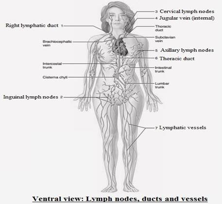
1. Right lymphatic duct:
It is a short vessel which receives the lymph from the right side of the body (mainly head, thorax and neck, right side of the heart, the right lung, right arm, and convex liver surface. This duct discharges at its junction into the right subclavian vein with the help of right internal jugular vein.
2. Inguinal lymph nodes:
These are the lymph nodes present in the human groin. These are in the femoral triangle of the inguinal region. These are assembled into the deep and superficial lymph nodes.
3. Cervical lymph nodes:
These are simply small lymph nodes which are present in the neck region. These are present all over the body. These play a significant role in the immune system.
4. The jugular vein:
It is a significant body part as it drains the deoxygenated blood. It transports the blood from the neck and the head.
5. Axillary lymph node:
It is a lymph node present in the armpit region which drains lymph from the breast and adjacent regions.
6. Thoracic duct:
It is the chief vessel of the lymphatic system. It passes upwards in front of the spine. It drains the fluidnear the neck base into the left innominate vein.
The lymph node is a small bean-shaped structure. It is in continuation of the lymphatic vessels. It is a part of the immune system of the body. Lymph nodes help in the filtration of substances present in the lymphatic fluid.
Want to see more full solutions like this?
Chapter 42 Solutions
Laboratory Manual for Holes Human Anatomy & Physiology Fetal Pig Version


- Ch.21 What causes patients infected with the yellow fever virus to turn yellow (jaundice)? A. low blood pressure and anemia B. excess leukocytes C. alteration of skin pigments D. liver damage in final stage of disease — What is the advantage for malarial parasites to grow and replicate in red blood cells? A. able to spread quickly B. able to avoid immune detection C. low oxygen environment for growth D. cooler area of the body for growth — Which microbe does not live part of its lifecycle outside humans? A. Toxoplasma gondii B. Cytomegalovirus C. Francisella tularensis D. Plasmodium falciparum — explain your answer thoroughlyarrow_forwardCh.22 Streptococcus pneumoniae has a capsule to protect it from killing by alveolar macrophages, which kill bacteria by… A. cytokines B. antibodies C. complement D. phagocytosis — What fact about the influenza virus allows the dramatic antigenic shift that generates novel strains? A. very large size B. enveloped C. segmented genome D. over 100 genes — explain your answer thoroughlyarrow_forwardWhat is this?arrow_forward
- Molecular Biology A-C components of the question are corresponding to attached image labeled 1. D component of the question is corresponding to attached image labeled 2. For a eukaryotic mRNA, the sequences is as follows where AUGrepresents the start codon, the yellow is the Kozak sequence and (XXX) just represents any codonfor an amino acid (no stop codons here). G-cap and polyA tail are not shown A. How long is the peptide produced?B. What is the function (a sentence) of the UAA highlighted in blue?C. If the sequence highlighted in blue were changed from UAA to UAG, how would that affecttranslation? D. (1) The sequence highlighted in yellow above is moved to a new position indicated below. Howwould that affect translation? (2) How long would be the protein produced from this new mRNA? Thank youarrow_forwardMolecular Biology Question Explain why the cell doesn’t need 61 tRNAs (one for each codon). Please help. Thank youarrow_forwardMolecular Biology You discover a disease causing mutation (indicated by the arrow) that alters splicing of its mRNA. This mutation (a base substitution in the splicing sequence) eliminates a 3’ splice site resulting in the inclusion of the second intron (I2) in the final mRNA. We are going to pretend that this intron is short having only 15 nucleotides (most introns are much longer so this is just to make things simple) with the following sequence shown below in bold. The ( ) indicate the reading frames in the exons; the included intron 2 sequences are in bold. A. Would you expected this change to be harmful? ExplainB. If you were to do gene therapy to fix this problem, briefly explain what type of gene therapy youwould use to correct this. Please help. Thank youarrow_forward
- Molecular Biology Question Please help. Thank you Explain what is meant by the term “defective virus.” Explain how a defective virus is able to replicate.arrow_forwardMolecular Biology Explain why changing the codon GGG to GGA should not be harmful. Please help . Thank youarrow_forwardStage Percent Time in Hours Interphase .60 14.4 Prophase .20 4.8 Metaphase .10 2.4 Anaphase .06 1.44 Telophase .03 .72 Cytukinesis .01 .24 Can you summarize the results in the chart and explain which phases are faster and why the slower ones are slow?arrow_forward
- Can you circle a cell in the different stages of mitosis? 1.prophase 2.metaphase 3.anaphase 4.telophase 5.cytokinesisarrow_forwardWhich microbe does not live part of its lifecycle outside humans? A. Toxoplasma gondii B. Cytomegalovirus C. Francisella tularensis D. Plasmodium falciparum explain your answer thoroughly.arrow_forwardSelect all of the following that the ablation (knockout) or ectopoic expression (gain of function) of Hox can contribute to. Another set of wings in the fruit fly, duplication of fingernails, ectopic ears in mice, excess feathers in duck/quail chimeras, and homeosis of segment 2 to jaw in Hox2a mutantsarrow_forward
 Medical Terminology for Health Professions, Spira...Health & NutritionISBN:9781305634350Author:Ann Ehrlich, Carol L. Schroeder, Laura Ehrlich, Katrina A. SchroederPublisher:Cengage Learning
 Principles Of Radiographic Imaging: An Art And A ...Health & NutritionISBN:9781337711067Author:Richard R. Carlton, Arlene M. Adler, Vesna BalacPublisher:Cengage Learning
 Nutrition Through The Life CycleHealth & NutritionISBN:9781337919333Author:Brown, Judith E.Publisher:Cengage Learning,