Concept explainers
Videos
Explain how connective tissues differ from epithelial tissues in structure and function.
To review:
The difference between connective tissues and epithelial tissues, based on their structure and function.
Introduction:
Tissues are a group of cells that have a specific function. In a living organism, tissues are of four types, namely, epithelial tissue, connective tissue, muscle tissue, and connective tissue. The function and structure of these tissues vary from each other.
Explanation of Solution
The difference between connective tissues and epithelial tissues is given in the following table:
| Connective tissue | Epithelial tissue |
| This group of tissues connects different tissues with each other. | Epithelial tissue is the most abundant tissue as it covers the cavities and the surface of the body. |
| They have an extracellular matrix (ECM) where cells are scattered. Fibers and ground substances, are important structural parts of ECM. | The cells are arranged in a sheet-like manner with little ECM. There are no blood vessels in epithelial tissues. |
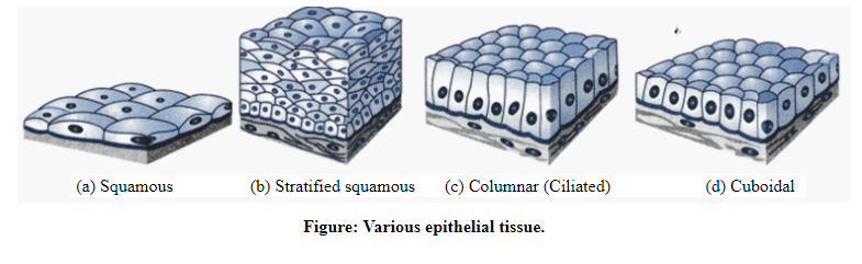
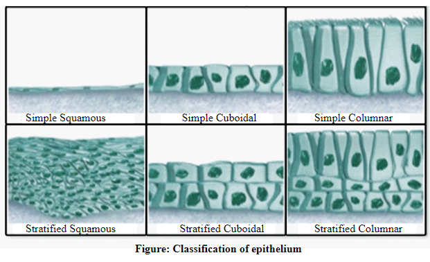
| Connective tissues are classified into proper connective tissue and specialized connective tissue. | Based on the tissue layer, epithelial tissues can be classified into simple and stratified. Simple epithelia have a single layer, whereas stratified epithelia have more than one layer. |
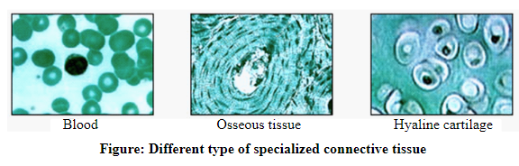
| Specialized connective tissues are cartilage, bone, and blood. Proper connective tissues are again classified into loose connective tissues, dense connective tissues, reticular tissues, and adipose tissues. | They are seen in three different principal shapes, namely, squamous, columnar, and cuboidal. |
| Blood is an example of a connective tissue. It transports nutrients and oxygen to various organs. Connective tissues provide support to our skeletal system. An example of a connective tissue that provides support is cartilage and dense connective tissue. | Epithelial tissues act as a barrier against mechanical and thermal injury. The best example is skin. |
The diagrams showing the structures of different types of connective and epithelial tissues are shown below:
Thus, it can be concluded that the two tissue classifications, namely, epitheliumand connective tissues, play a major role in the defense mechanism of the body. Based on their composition, they can also be further classified into different types.
Want to see more full solutions like this?
Chapter 4 Solutions
Human Anatomy & Physiology, Books a la Carte Edition (2nd Edition)


- What is this?arrow_forwardMolecular Biology A-C components of the question are corresponding to attached image labeled 1. D component of the question is corresponding to attached image labeled 2. For a eukaryotic mRNA, the sequences is as follows where AUGrepresents the start codon, the yellow is the Kozak sequence and (XXX) just represents any codonfor an amino acid (no stop codons here). G-cap and polyA tail are not shown A. How long is the peptide produced?B. What is the function (a sentence) of the UAA highlighted in blue?C. If the sequence highlighted in blue were changed from UAA to UAG, how would that affecttranslation? D. (1) The sequence highlighted in yellow above is moved to a new position indicated below. Howwould that affect translation? (2) How long would be the protein produced from this new mRNA? Thank youarrow_forwardMolecular Biology Question Explain why the cell doesn’t need 61 tRNAs (one for each codon). Please help. Thank youarrow_forward
- Molecular Biology You discover a disease causing mutation (indicated by the arrow) that alters splicing of its mRNA. This mutation (a base substitution in the splicing sequence) eliminates a 3’ splice site resulting in the inclusion of the second intron (I2) in the final mRNA. We are going to pretend that this intron is short having only 15 nucleotides (most introns are much longer so this is just to make things simple) with the following sequence shown below in bold. The ( ) indicate the reading frames in the exons; the included intron 2 sequences are in bold. A. Would you expected this change to be harmful? ExplainB. If you were to do gene therapy to fix this problem, briefly explain what type of gene therapy youwould use to correct this. Please help. Thank youarrow_forwardMolecular Biology Question Please help. Thank you Explain what is meant by the term “defective virus.” Explain how a defective virus is able to replicate.arrow_forwardMolecular Biology Explain why changing the codon GGG to GGA should not be harmful. Please help . Thank youarrow_forward
- Stage Percent Time in Hours Interphase .60 14.4 Prophase .20 4.8 Metaphase .10 2.4 Anaphase .06 1.44 Telophase .03 .72 Cytukinesis .01 .24 Can you summarize the results in the chart and explain which phases are faster and why the slower ones are slow?arrow_forwardCan you circle a cell in the different stages of mitosis? 1.prophase 2.metaphase 3.anaphase 4.telophase 5.cytokinesisarrow_forwardWhich microbe does not live part of its lifecycle outside humans? A. Toxoplasma gondii B. Cytomegalovirus C. Francisella tularensis D. Plasmodium falciparum explain your answer thoroughly.arrow_forward
- Select all of the following that the ablation (knockout) or ectopoic expression (gain of function) of Hox can contribute to. Another set of wings in the fruit fly, duplication of fingernails, ectopic ears in mice, excess feathers in duck/quail chimeras, and homeosis of segment 2 to jaw in Hox2a mutantsarrow_forwardSelect all of the following that changes in the MC1R gene can lead to: Changes in spots/stripes in lizards, changes in coat coloration in mice, ectopic ear formation in Siberian hamsters, and red hair in humansarrow_forwardPleiotropic genes are genes that (blank) Cause a swapping of organs/structures, are the result of duplicated sets of chromosomes, never produce protein products, and have more than one purpose/functionarrow_forward
 Human Biology (MindTap Course List)BiologyISBN:9781305112100Author:Cecie Starr, Beverly McMillanPublisher:Cengage Learning
 Human Physiology: From Cells to Systems (MindTap ...BiologyISBN:9781285866932Author:Lauralee SherwoodPublisher:Cengage Learning

 Biology: The Unity and Diversity of Life (MindTap...BiologyISBN:9781305073951Author:Cecie Starr, Ralph Taggart, Christine Evers, Lisa StarrPublisher:Cengage Learning