
Videos
(a)
To identify: The interphase, each phase of mitosis and cytokinesis in the figure 4.4(a)-(e).
Introduction: The cell division is the process of cycle cycle in which the parent cell divided into two or more daughter cells. The cell divison is needed for growth of the individual, wound healing and replacement of old and dying cells. The duration of cell division differs on the type of the cell. The somatic cell divison consists of three phases namely interphase, mitotic phase and cytokinesis.
(a)
Answer to Problem 1.1BGL
Pictorial representation: The phase of cell cycle given in the figure 4.4 (a) is identified as interphase.
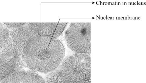
Fig 1: The interphase of somatic cell division.
Explanation of Solution
The interphase is the longest part of the cell cycle. During this phase the cell grow and replicates its DNA before moving into the mitosis. The interphase consist of three stages anmely G1 phase, S phase and G2 phase. The G1 phase is the first gap pahse in which the cell grows larger and organeeles are copied. The S phase is the synthetic phase in which the DNA of the cell replicates to produce another copy of DNA. The G2 phase is the second gap pahse in which the cells makes proteins and reorganise its contents before it enters into the mitosis.
(b)
To identify: The interphase, each phase of mitosis and cytokinesis in the figure 4.4(a)-(e).
Introduction: The cell division is the process of cycle cycle in which the parent cell divided into two or more daughter cells. The cell divison is needed for growth of the individual, wound healing and replacement of old and dying cells. The duration of cell division differs on the type of the cell. The somatic cell divison consists of three phases namely interphase, mitotic phase and cytokinesis.
(b)
Answer to Problem 1.1BGL
Pictorial representation: The phase of cell cycle given in the figure 4.4 (b) is identified as prophase.
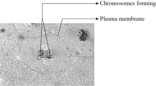
Fig 2: The prophase of somatic cell division.
Explanation of Solution
The mitotic phase of the cell division is called as M phase. This phase consists of four stages namely prophase, metaphase, anaphase and telophase. The prophase is the first phase of cell division. During this phase, the chromosomes becomes visible, the nucleolus disappears, the chromatin condenses, the mitotic spindle forms and the nuclear envelope disappears.
(c)
To identify: The interphase, each phase of mitosis and cytokinesis in the figure 4.4(a)-(e).
Introduction: The cell division is the process of cycle cycle in which the parent cell divided into two or more daughter cells. The cell divison is needed for growth of the individual, wound healing and replacement of old and dying cells. The duration of cell division differs on the type of the cell. The somatic cell divison consists of three phases namely interphase, mitotic phase and cytokinesis.
(c)
Answer to Problem 1.1BGL
Pictorial representation: The phase of cell cycle given in the figure 4.4 (c) is identified as metaphase.
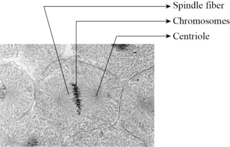
Fig 3: The metaphase of somatic cell division.
Explanation of Solution
The metaphase is the second phase of mitosis. At the start of metaphase the chromosomes are randomly arranged within the nucleus. The microtubules connects to each chromosome. Then the chromosomes are aligned in the middle of the cell with the sister chromatids of each chromosome on either side of the metaphase plate. The sister chromatids contains cohensin proteins which are broken after passing the mitotic check point to ensure alignment of chromosomes at the metaphase plate. This then acrtivates the anaphase-promoting complex which leads to end of metaphase.
(d)
To identify: The interphase, each phase of mitosis and cytokinesis in the figure 4.4(a)-(e).
Introduction: The cell division is the process of cycle cycle in which the parent cell divided into two or more daughter cells. The cell divison is needed for growth of the individual, wound healing and replacement of old and dying cells. The duration of cell division differs on the type of the cell. The somatic cell divison consists of three phases namely interphase, mitotic phase and cytokinesis.
(d)
Answer to Problem 1.1BGL
Pictorial representation: The phase of cell cycle given in the figure 4.4 (d) is identified as anaphase.
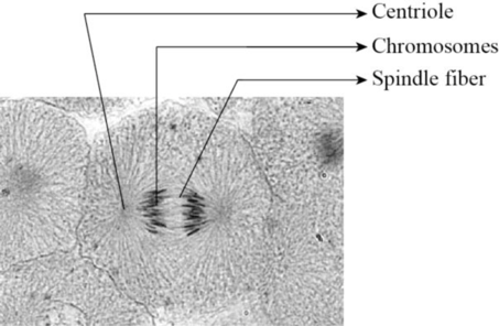
Fig 4: The anaphase of somatic cell division.
Explanation of Solution
The anaphase of cell cycle begins with the formation of anaphase promoting complex. This complex degrades the protein called securin which inhibits the separase. The separase is the protein that separates the cohesion protein that helds the chromatids together. As the result of this process the chromatids becomes apart from each other.
(e)
To identify: The interphase, each phase of mitosis and cytokinesis in the figure 4.4(a)-(e).
Introduction: The cell division is the process of cycle cycle in which the parent cell divided into two or more daughter cells. The cell divison is needed for growth of the individual, wound healing and replacement of old and dying cells. The duration of cell division differs on the type of the cell. The somatic cell divison consists of three phases namely interphase, mitotic phase and cytokinesis.
(e)
Answer to Problem 1.1BGL
Pictorial representation: The phase of cell cycle given in the figure 4.4 (e) is identified as telophase.
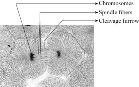
Fig 5: The telophase of somatic cell division.
Explanation of Solution
The telophase is the final stage of cell cycle. During this phase, the spindle fibers underoes depolymerisation that separstes the sister chromatids. The separate chromosomes are released from tighltly bound structure. Then the nuclear envelops reforms around the new nuclei in each half of the dividing cells.
Want to see more full solutions like this?
Chapter 4 Solutions
Laboratory Manual for Anatomy and Physiology, 6e Loose-Leaf Print Companion with WileyPLUS Blackboard Card Set


- “Gretchen” was a 68-pound canine who came to the VMTH as small animal surgery patient. She receivedacepromazine, 0.2 mg/kg from a 10 mg/ml solution and oxymorphone, 0.08 mg/kg from a 1 mg/ml solution before surgery.What are the mechanisms of action of acepromazine and oxymorphone? Why would they be given together?How many mg provide each dose and how many ml of each of these solutions were given?arrow_forwardAfter surgery, “Gretchen” was put on carprofen, 1 mg/pound bid (twice a day). The tablets come in 25, 75 and 100 mgsizes. Which size tablet would be appropriate?What is the mechanism of action of carprofen?An outpatient prescription was written for her so she would have enough for 10 days. How many tablets did she need?What information needs to be on her out-patient prescription?arrow_forwardJoden Koepp olor in chickens is due to incomplete dominance. BB = Black chicken, WW = White BLOOD TYPES Arhite chicken is In humans, Rh positive blood is dominant (R) over Rh negative blood (r). A man with type 0, Rh positive blood (whose mother had Rh negative blood), marries a woman with type AB, Rh negative blood. Several children were born. is? R R Genotypes Phenotypes RRR RR Rr Rr 4/16 RR R RR RK Rr Rr 4/16 rr 3/4 Rh posi 1/4 Rh negu 1/2 Rr rr rr rrrr 88 888 75 e genotype of the man? the genotype of the woman? The mother of the man had type AB blood.arrow_forward
- Please indentify the unknown organismarrow_forward5G JA ATTC 3 3 CTIA A1G5 5 GAAT I I3 3 CTIA AA5 Fig. 5-3: The Eco restriction site (left) would be cleaved at the locations indicated by the arrows. However, a SNP in the position shown in gray (right) would prevent cleavage at this site by EcoRI One of the SNPs in B. rapa is found within the Park14 locus and can be detected by RFLP analysis. The CT polymorphism is found in the intron of the Bra013780 gene found on Chromosome 1. The Park14 allele with the "C" in the SNP has two EcoRI sites and thus is cleaved twice by EcoRI If there is a "T" in that SNP, one of the EcoRI sites is altered, so the Park14 allele with the T in the SNP has only one EcoRI site (Fig. 5-3). Park14 allele with SNP(C) Park14 allele with SNPT) 839 EcoRI 1101 EcoRI 839 EcoRI Fig. 5.4: Schematic restriction maps of the two different Park14 alleles (1316 bp long) of B. rapa. Where on these maps is the CT SNP located? 90 The primers used to amplify the DNA at the Park14 locus (see Fig. 5 and Table 3 of Slankster et…arrow_forwardFrom your previous experiment, you found that this enhancer activates stripe 2 of eve expression. When you sequence this enhancer you find several binding sites for the gap gene, Giant. To test how Giant interacts with eve, you decide to remove all of the Giant binding sites from the eve enhancer. What results do you expect to see with respect to eve expression?arrow_forward
- What experiment could you do to see if the maternal gene, bicoid, is sufficient to form anterior fates?arrow_forwardYou’re curious about the effect that gap genes have on the pair-rule gene, evenskipped (eve), so you isolate and sequence each of the eve enhancers. You’re particularly interested in one of the enhancers, which is just upstream of the eve gene. Describe an experimental technique you would use to find out where this particular eve enhancer is active.arrow_forwardFor short answer questions, write your answers on the line provided. To the right is the mRNA codon table to use as needed throughout the exam. First letter U บบบ U CA UUCPhe UUA UCU Phe UCC UUG Leu CUU UAU. G U UAC TV UGCys UAA Stop UGA Stop A UAG Stop UGG Trp Ser UCA UCG CCU] 0 CUC CUA CCC CAC CAU His CGU CGC Leu Pro CCA CAA Gin CGA Arg CUG CCG CAG CGG AUU ACU AAU T AUC lle A 1 ACC Thr AUA ACA AUG Mot ACG AGG Arg GUU GCU GUC GCC G Val Ala GAC Asp GGU GGC GUA GUG GCA GCG GAA GGA Gly Glu GAGJ GGG AACASH AGU Ser AAA1 AAG Lys GAU AGA CAL CALUCAO CAO G Third letter 1. (+7) Use the table below to answer the questions; use the codon table above to assist you. The promoter sequence of DNA is on the LEFT. You do not need to fill in the entire table. Assume we are in the middle of a gene sequence (no need to find a start codon). DNA 1 DNA 2 mRNA tRNA Polypeptide C Val G C. T A C a. On which strand of DNA is the template strand (DNA 1 or 2)?_ b. On which side of the mRNA is the 5' end (left or…arrow_forward
 Human Anatomy & Physiology (11th Edition)BiologyISBN:9780134580999Author:Elaine N. Marieb, Katja N. HoehnPublisher:PEARSON
 Biology 2eBiologyISBN:9781947172517Author:Matthew Douglas, Jung Choi, Mary Ann ClarkPublisher:OpenStax
 Anatomy & PhysiologyBiologyISBN:9781259398629Author:McKinley, Michael P., O'loughlin, Valerie Dean, Bidle, Theresa StouterPublisher:Mcgraw Hill Education,
 Molecular Biology of the Cell (Sixth Edition)BiologyISBN:9780815344322Author:Bruce Alberts, Alexander D. Johnson, Julian Lewis, David Morgan, Martin Raff, Keith Roberts, Peter WalterPublisher:W. W. Norton & Company
 Laboratory Manual For Human Anatomy & PhysiologyBiologyISBN:9781260159363Author:Martin, Terry R., Prentice-craver, CynthiaPublisher:McGraw-Hill Publishing Co.
 Inquiry Into Life (16th Edition)BiologyISBN:9781260231700Author:Sylvia S. Mader, Michael WindelspechtPublisher:McGraw Hill Education