Videos
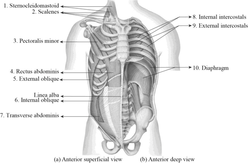
To label: The structures in Figure 33.1.
Introduction: The muscles that are involved in the process of breathing are referred to as the respiratory muscles. These muscles contribute to the inhalation and exhalation processes by aiding the contraction and expansion of the thoracic cavity. The diaphragm, internal and external intercostals, pectoralis minors, scalenes, sternocleidomastoids, and abdominal muscles are the primary muscles of the respiration.
Answer to Problem 1.1BGL
Pictorial representation:
Explanation of Solution
1. Sternocleidomastoid: A strap-like muscle that is situated on the lateral and the anterior portion of the neck is termed as the sternocleidomastoid. These muscles are involved in the respiration process by working along with the scalene muscles. These muscles are involved in raising the sternum during the forced inspiration while breathing.
2. Scalenes: A group of three pairs of muscles situated in the lateral region of the neck is referred to as the scalenes. The three pairs of these muscles include the anterior, middle, and posterior scalenes. The primary action of the scalene muscles is the elevation of the first and second ribs.
3. Pectoralis minor: The pectoralis minor is a flat, thin-shaped muscle located beneath the pectoralis major. This muscle elevates from the 3rd, 4th, and 5th ribs on each side of the rib cage. From these locations, the pectoralis minor muscle extends up to the chest and gets inserted into the coracoid process of the shoulder blade. The main function of this muscle is to expand the rib cage for aiding inhalation. This would help to expand the lungs and hold the breath for a long period of time.
4. Rectus abdominis: The rectus abdominis is a long strap muscle that begins at the pubic bone and ends at the sternum. They are the midline abdominal muscles that are situated between the inguinal ligament and the sternum. The main function of these muscles is flexion of the vertebral column and compression of the abdomen. It plays an essential role in respiration with exhalation during exercises.
5. External oblique: The external oblique muscles are the largest and outermost abdominal muscles. They are wide thin muscles that run diagonally downward each side of the abdomen. These muscles help to rotate the trunk. They pull the chest downward and compress the abdominal cavity.
6. Internal oblique: The internal oblique muscles are located beneath the external obliques. The fibers of these muscles run obliquely up toward the linea alba (band of the connective tissue, which runs up the midline of the abdomen). The main action of the internal obliques includes the flexion, lateral rotation, and lateral flexion of the trunk.
7. Transverse abdominis: The transverse abdominis is a muscle layer that is present in the anterior and lateral side of the abdominal wall. They are situated deep within the internal oblique muscles. This muscle is also termed as the transversus abdominis and transverse abdominal muscle (TVA). The main function of this muscle is to activate the core muscles and to stabilize the low back and pelvis prior to the body movement.
8. Internal intercostals: A group of 22 pairs of tiny skeletal muscles that are located in between the skeletal muscles are referred to as the internal intercostals. They play a vital role in the chest movement during the process of breathing. These muscles are involved in the process of forced exhalation by depressing the ribs.
9. External intercostals: A group of muscles that are located in between the outside of the ribs is referred to as the external intercostals. They play a vital role in the chest movement during the process of breathing. These muscles are involved in the process of forced and quiet inhalation of air. The primary action of these muscles is to elevation or raising and expansion of the ribs.
10. Diaphragm: A thin skeletal muscle that is situated at the base of the chest is referred to as a diaphragm. It separates the abdominal cavity from the chest. It plays a vital role in the process of respiration. It is involved in pulling the air into the lungs by creating a vacuum effect. During inhalation, the diaphragm is contracted and gets flattened. During exhalation, the diaphragm gets relaxed and pushes the air out of the lungs.
Want to see more full solutions like this?
Chapter 33 Solutions
Laboratory Manual for Anatomy and Physiology, 6e Loose-Leaf Print Companion


- What is this?arrow_forwardMolecular Biology A-C components of the question are corresponding to attached image labeled 1. D component of the question is corresponding to attached image labeled 2. For a eukaryotic mRNA, the sequences is as follows where AUGrepresents the start codon, the yellow is the Kozak sequence and (XXX) just represents any codonfor an amino acid (no stop codons here). G-cap and polyA tail are not shown A. How long is the peptide produced?B. What is the function (a sentence) of the UAA highlighted in blue?C. If the sequence highlighted in blue were changed from UAA to UAG, how would that affecttranslation? D. (1) The sequence highlighted in yellow above is moved to a new position indicated below. Howwould that affect translation? (2) How long would be the protein produced from this new mRNA? Thank youarrow_forwardMolecular Biology Question Explain why the cell doesn’t need 61 tRNAs (one for each codon). Please help. Thank youarrow_forward
- Molecular Biology You discover a disease causing mutation (indicated by the arrow) that alters splicing of its mRNA. This mutation (a base substitution in the splicing sequence) eliminates a 3’ splice site resulting in the inclusion of the second intron (I2) in the final mRNA. We are going to pretend that this intron is short having only 15 nucleotides (most introns are much longer so this is just to make things simple) with the following sequence shown below in bold. The ( ) indicate the reading frames in the exons; the included intron 2 sequences are in bold. A. Would you expected this change to be harmful? ExplainB. If you were to do gene therapy to fix this problem, briefly explain what type of gene therapy youwould use to correct this. Please help. Thank youarrow_forwardMolecular Biology Question Please help. Thank you Explain what is meant by the term “defective virus.” Explain how a defective virus is able to replicate.arrow_forwardMolecular Biology Explain why changing the codon GGG to GGA should not be harmful. Please help . Thank youarrow_forward
- Stage Percent Time in Hours Interphase .60 14.4 Prophase .20 4.8 Metaphase .10 2.4 Anaphase .06 1.44 Telophase .03 .72 Cytukinesis .01 .24 Can you summarize the results in the chart and explain which phases are faster and why the slower ones are slow?arrow_forwardCan you circle a cell in the different stages of mitosis? 1.prophase 2.metaphase 3.anaphase 4.telophase 5.cytokinesisarrow_forwardWhich microbe does not live part of its lifecycle outside humans? A. Toxoplasma gondii B. Cytomegalovirus C. Francisella tularensis D. Plasmodium falciparum explain your answer thoroughly.arrow_forward
- Select all of the following that the ablation (knockout) or ectopoic expression (gain of function) of Hox can contribute to. Another set of wings in the fruit fly, duplication of fingernails, ectopic ears in mice, excess feathers in duck/quail chimeras, and homeosis of segment 2 to jaw in Hox2a mutantsarrow_forwardSelect all of the following that changes in the MC1R gene can lead to: Changes in spots/stripes in lizards, changes in coat coloration in mice, ectopic ear formation in Siberian hamsters, and red hair in humansarrow_forwardPleiotropic genes are genes that (blank) Cause a swapping of organs/structures, are the result of duplicated sets of chromosomes, never produce protein products, and have more than one purpose/functionarrow_forward
 Human Anatomy & Physiology (11th Edition)BiologyISBN:9780134580999Author:Elaine N. Marieb, Katja N. HoehnPublisher:PEARSON
 Biology 2eBiologyISBN:9781947172517Author:Matthew Douglas, Jung Choi, Mary Ann ClarkPublisher:OpenStax
 Anatomy & PhysiologyBiologyISBN:9781259398629Author:McKinley, Michael P., O'loughlin, Valerie Dean, Bidle, Theresa StouterPublisher:Mcgraw Hill Education,
 Molecular Biology of the Cell (Sixth Edition)BiologyISBN:9780815344322Author:Bruce Alberts, Alexander D. Johnson, Julian Lewis, David Morgan, Martin Raff, Keith Roberts, Peter WalterPublisher:W. W. Norton & Company
 Laboratory Manual For Human Anatomy & PhysiologyBiologyISBN:9781260159363Author:Martin, Terry R., Prentice-craver, CynthiaPublisher:McGraw-Hill Publishing Co.
 Inquiry Into Life (16th Edition)BiologyISBN:9781260231700Author:Sylvia S. Mader, Michael WindelspechtPublisher:McGraw Hill Education