BIOLOGY:CONCEPTS+APPL.(LOOSELEAF)
10th Edition
ISBN: 9781305967359
Author: STARR
Publisher: CENGAGE L
expand_more
expand_more
format_list_bulleted
Concept explainers
Videos
Textbook Question
Chapter 28, Problem 4CT
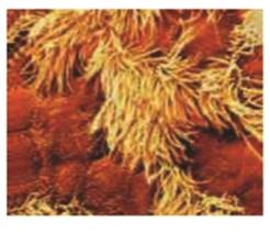
The micrograph to the left shows cells from the lining of an airway leading to the lungs. The gold cells are ciliated and the darker brown ones secrete mucus. What type of tissue is this? How can you tell?
Expert Solution & Answer
Want to see the full answer?
Check out a sample textbook solution
Students have asked these similar questions
what key characteristics would you look for when identifying microbes?
If you had an unknown microbe, what steps would you take to determine what type of microbe (e.g., fungi, bacteria, virus) it is? Are there particular characteristics you would search for? Explain.
avorite Contact
avorite Contact
favorite Contact
୫
Recant Contacts
Keypad
Messages
Pairing
ง
107.5
NE
Controls
Media Apps Radio
Nav Phone
SCREEN
OFF
Safari File Edit View History Bookmarks Window Help
newconnect.mheducation.com
M Sign in...
S The Im...
QFri May 9 9:23 PM
w The Im...
My first....
Topic:
Mi Kimberl
M Yeast F
Connection lost! You are not connected to internet
Sigh in...
Sign in...
The Im...
S Workin...
The Im.
INTRODUCTION
LABORATORY SIMULATION
Tube 1
Fructose)
esc
- X
Tube 2
(Glucose)
Tube 3
(Sucrose)
Tube 4
(Starch)
Tube 5
(Water)
CO₂ Bubble Height (mm)
How to Measure
92
3
5
6
METHODS
RESET
#3
W
E
80
A
S
D
9
02
1
2
3
5
2
MY NOTES
LAB DATA
SHOW LABELS
%
5
T
M dtv
96
J:
ப
27
כ
00
alt
A
DII
FB
G
H
J
K
PHASE 4:
Measure gas bubble
Complete the following steps:
Select ruler and place next to tube
1. Measure starting height of gas
bubble in respirometer 1. Record in
Lab Data
Repeat measurement for tubes 2-5
by selecting ruler and move next to
each tube. Record each in Lab
Data…
Chapter 28 Solutions
BIOLOGY:CONCEPTS+APPL.(LOOSELEAF)
Ch. 28 - _______ tissues are sheetlike with one free...Ch. 28 - _________ allow cardiac muscle cells to contract...Ch. 28 - Glands are derived from ________ tissue. a....Ch. 28 - Most ________ have many collagen and elastin...Ch. 28 - ________ is mostly plasma. a. Adipose tissue c....Ch. 28 - Prob. 6SACh. 28 - Cells of ________ can shorten contract. a....Ch. 28 - Prob. 8SACh. 28 - ________ detects and integrates information about...Ch. 28 - Prob. 10SA
Ch. 28 - Prob. 11SACh. 28 - Prob. 12SACh. 28 - Prob. 13SACh. 28 - Prob. 14SACh. 28 - Match the terms with the most suitable...Ch. 28 - Many people oppose the use of animals for testing...Ch. 28 - Prob. 2CTCh. 28 - Each level of biological organization has emergent...Ch. 28 - The micrograph to the left shows cells from the...
Knowledge Booster


Learn more about
Need a deep-dive on the concept behind this application? Look no further. Learn more about this topic, biology and related others by exploring similar questions and additional content below.Similar questions
- Ch.23 How is Salmonella able to cross from the intestines into the blood? A. it is so small that it can squeeze between intestinal cells B. it secretes a toxin that induces its uptake into intestinal epithelial cells C. it secretes enzymes that create perforations in the intestine D. it can get into the blood only if the bacteria are deposited directly there, that is, through a puncture — Which virus is associated with liver cancer? A. hepatitis A B. hepatitis B C. hepatitis C D. both hepatitis B and C — explain your answer thoroughlyarrow_forwardCh.21 What causes patients infected with the yellow fever virus to turn yellow (jaundice)? A. low blood pressure and anemia B. excess leukocytes C. alteration of skin pigments D. liver damage in final stage of disease — What is the advantage for malarial parasites to grow and replicate in red blood cells? A. able to spread quickly B. able to avoid immune detection C. low oxygen environment for growth D. cooler area of the body for growth — Which microbe does not live part of its lifecycle outside humans? A. Toxoplasma gondii B. Cytomegalovirus C. Francisella tularensis D. Plasmodium falciparum — explain your answer thoroughlyarrow_forwardCh.22 Streptococcus pneumoniae has a capsule to protect it from killing by alveolar macrophages, which kill bacteria by… A. cytokines B. antibodies C. complement D. phagocytosis — What fact about the influenza virus allows the dramatic antigenic shift that generates novel strains? A. very large size B. enveloped C. segmented genome D. over 100 genes — explain your answer thoroughlyarrow_forward
- What is this?arrow_forwardMolecular Biology A-C components of the question are corresponding to attached image labeled 1. D component of the question is corresponding to attached image labeled 2. For a eukaryotic mRNA, the sequences is as follows where AUGrepresents the start codon, the yellow is the Kozak sequence and (XXX) just represents any codonfor an amino acid (no stop codons here). G-cap and polyA tail are not shown A. How long is the peptide produced?B. What is the function (a sentence) of the UAA highlighted in blue?C. If the sequence highlighted in blue were changed from UAA to UAG, how would that affecttranslation? D. (1) The sequence highlighted in yellow above is moved to a new position indicated below. Howwould that affect translation? (2) How long would be the protein produced from this new mRNA? Thank youarrow_forwardMolecular Biology Question Explain why the cell doesn’t need 61 tRNAs (one for each codon). Please help. Thank youarrow_forward
- Molecular Biology You discover a disease causing mutation (indicated by the arrow) that alters splicing of its mRNA. This mutation (a base substitution in the splicing sequence) eliminates a 3’ splice site resulting in the inclusion of the second intron (I2) in the final mRNA. We are going to pretend that this intron is short having only 15 nucleotides (most introns are much longer so this is just to make things simple) with the following sequence shown below in bold. The ( ) indicate the reading frames in the exons; the included intron 2 sequences are in bold. A. Would you expected this change to be harmful? ExplainB. If you were to do gene therapy to fix this problem, briefly explain what type of gene therapy youwould use to correct this. Please help. Thank youarrow_forwardMolecular Biology Question Please help. Thank you Explain what is meant by the term “defective virus.” Explain how a defective virus is able to replicate.arrow_forwardMolecular Biology Explain why changing the codon GGG to GGA should not be harmful. Please help . Thank youarrow_forward
- Stage Percent Time in Hours Interphase .60 14.4 Prophase .20 4.8 Metaphase .10 2.4 Anaphase .06 1.44 Telophase .03 .72 Cytukinesis .01 .24 Can you summarize the results in the chart and explain which phases are faster and why the slower ones are slow?arrow_forwardCan you circle a cell in the different stages of mitosis? 1.prophase 2.metaphase 3.anaphase 4.telophase 5.cytokinesisarrow_forwardWhich microbe does not live part of its lifecycle outside humans? A. Toxoplasma gondii B. Cytomegalovirus C. Francisella tularensis D. Plasmodium falciparum explain your answer thoroughly.arrow_forward
arrow_back_ios
SEE MORE QUESTIONS
arrow_forward_ios
Recommended textbooks for you

 Biology 2eBiologyISBN:9781947172517Author:Matthew Douglas, Jung Choi, Mary Ann ClarkPublisher:OpenStax
 Biology: The Unity and Diversity of Life (MindTap...BiologyISBN:9781305073951Author:Cecie Starr, Ralph Taggart, Christine Evers, Lisa StarrPublisher:Cengage Learning
 Human Biology (MindTap Course List)BiologyISBN:9781305112100Author:Cecie Starr, Beverly McMillanPublisher:Cengage Learning
Biology 2e
Biology
ISBN:9781947172517
Author:Matthew Douglas, Jung Choi, Mary Ann Clark
Publisher:OpenStax
Biology: The Unity and Diversity of Life (MindTap...
Biology
ISBN:9781305073951
Author:Cecie Starr, Ralph Taggart, Christine Evers, Lisa Starr
Publisher:Cengage Learning
Human Biology (MindTap Course List)
Biology
ISBN:9781305112100
Author:Cecie Starr, Beverly McMillan
Publisher:Cengage Learning
Types of Human Body Tissue; Author: MooMooMath and Science;https://www.youtube.com/watch?v=O0ZvbPak4ck;License: Standard YouTube License, CC-BY