Concept explainers
Videos
(a)
To label: The heart structures in Figure 27.2(a)
Introduction: The heart is the muscular organ that weighs about the size of the fist. The main function of the heart is to transport the oxygenated blood to all parts of the body. It consists of four chambers namely right atrium, left atrium, right ventricle, and left ventricle. The blood vessels called arteries and veins are involved in the transport of blood to the tissues and from tissues to the heart.
(a)
Answer to Problem 1.1BGL
Pictorial representation:
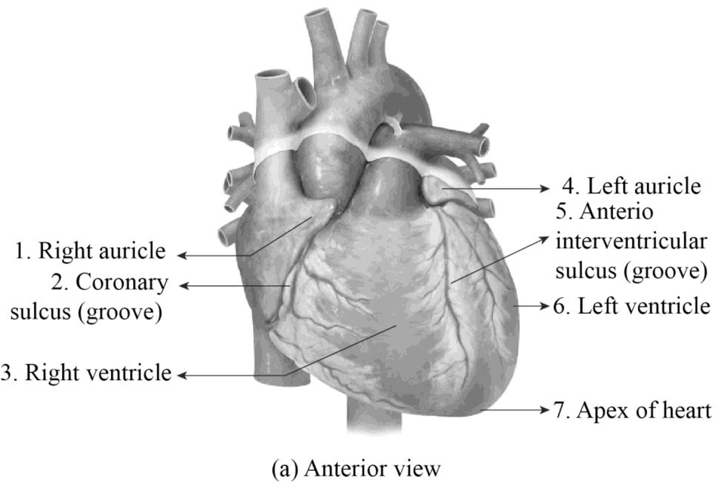
Fig 1: The anterior view of the heart.
Explanation of Solution
The structures of the anterior view of the heart is reviewed as follows:
Right auricle: It is the muscular cone shaped pouch that is attached to the right atrium of the heart. It is also called as right atrial appendage.
Cononary sulcus: It is the structure that separates the atrium of the heart from the ventricles. There are two coronary sulcus present in the heart namely right coronary sulcus and left coronary sulcus.
Right ventricle: It is one of the four heart chambers. It plays an important role in carrying the deoxygenated blood from the heart to the lungs.
Left auricle: It is the muscular cone shaped pouch that is attached to the anterior surface of the left atrium of the heart. It functions in increasing the pumping capacity of the left atrium.
Anterior interventricular sulcus: The heart consists of two interventricular sulcus namely anterior interventricular sulcus and posterior interventricular sulcus. It plays an important role in separating the ventricles of the heart.
Left ventricle: It is one of the four heart chambers. It plays an important role in carrying the oxygenated blood to all parts of the body.
Apex of heart: It is present in the left side of the heart between the fourth and fifth ribs. It consists of the left ventricle. It plays an important role in regulating the contraction of the ventricles.
(b)
To label: The heart structures in Figure 27.2(b)
Introduction: The heart is the muscular organ that weighs about the size of the fist. The main function of the heart is to transport the oxygenated blood to all parts of the body. It consists of four chambers namely right atrium, left atrium, right ventricle, and left ventricle. The blood vessels called arteries and veins are involved in the transport of blood to the tissues and from tissues to the heart.
(b)
Answer to Problem 1.1BGL
Pictorial representation:
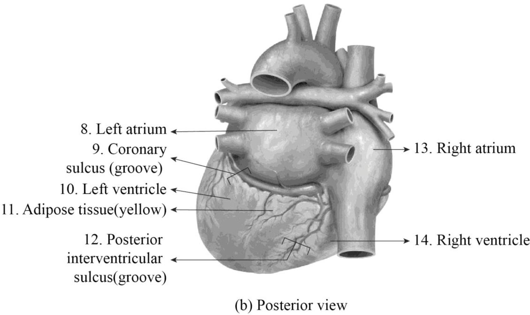
Fig 2: The posterior view of the heart.
Explanation of Solution
The structures of the posterior view of the heart is reviewed as follows:
Left atrium: It is one of the four heart chambers. The oxygenated blood from the lungs enters into the left atrium.
Coronary sulcus: It is the structure that separates the atrium of the heart from the ventricles. There are two coronary sulcus present in the heart namely right coronary sulcus and left coronary sulcus.
Left ventricle: It is one of the four heart chambers. It plays an important role in carrying the oxygenated blood to all parts of the body.
Adipose tissue: The adipose tissue that surrounds the heart plays an important role by acting as cushion and insulates the heart.
Posterior interventricular sulcus: The heart consists of two interventricular sulcus namely anterior interventricular sulcus and posterior interventricular sulcus. It plays an important role in separating the ventricles of the heart.
Right atrium: It is one of the four chambers of the heart that receives the deoxygenated blood from the vena cava and pumps to the right ventricle.
Right ventricle: It is one of the four chambers of the heart that sends blood to the lungs to get oxygenated through the pulmonary arteries.
Want to see more full solutions like this?
Chapter 27 Solutions
Laboratory Manual for Anatomy and Physiology, 6e Loose-Leaf Print Companion


- Please indentify the unknown organismarrow_forwardPlease indentify the unknown organismarrow_forward5G JA ATTC 3 3 CTIA A1G5 5 GAAT I I3 3 CTIA AA5 Fig. 5-3: The Eco restriction site (left) would be cleaved at the locations indicated by the arrows. However, a SNP in the position shown in gray (right) would prevent cleavage at this site by EcoRI One of the SNPs in B. rapa is found within the Park14 locus and can be detected by RFLP analysis. The CT polymorphism is found in the intron of the Bra013780 gene found on Chromosome 1. The Park14 allele with the "C" in the SNP has two EcoRI sites and thus is cleaved twice by EcoRI If there is a "T" in that SNP, one of the EcoRI sites is altered, so the Park14 allele with the T in the SNP has only one EcoRI site (Fig. 5-3). Park14 allele with SNP(C) Park14 allele with SNPT) 839 EcoRI 1101 EcoRI 839 EcoRI Fig. 5.4: Schematic restriction maps of the two different Park14 alleles (1316 bp long) of B. rapa. Where on these maps is the CT SNP located? 90 The primers used to amplify the DNA at the Park14 locus (see Fig. 5 and Table 3 of Slankster et…arrow_forward
- From your previous experiment, you found that this enhancer activates stripe 2 of eve expression. When you sequence this enhancer you find several binding sites for the gap gene, Giant. To test how Giant interacts with eve, you decide to remove all of the Giant binding sites from the eve enhancer. What results do you expect to see with respect to eve expression?arrow_forwardWhat experiment could you do to see if the maternal gene, bicoid, is sufficient to form anterior fates?arrow_forwardYou’re curious about the effect that gap genes have on the pair-rule gene, evenskipped (eve), so you isolate and sequence each of the eve enhancers. You’re particularly interested in one of the enhancers, which is just upstream of the eve gene. Describe an experimental technique you would use to find out where this particular eve enhancer is active.arrow_forward
- For short answer questions, write your answers on the line provided. To the right is the mRNA codon table to use as needed throughout the exam. First letter U บบบ U CA UUCPhe UUA UCU Phe UCC UUG Leu CUU UAU. G U UAC TV UGCys UAA Stop UGA Stop A UAG Stop UGG Trp Ser UCA UCG CCU] 0 CUC CUA CCC CAC CAU His CGU CGC Leu Pro CCA CAA Gin CGA Arg CUG CCG CAG CGG AUU ACU AAU T AUC lle A 1 ACC Thr AUA ACA AUG Mot ACG AGG Arg GUU GCU GUC GCC G Val Ala GAC Asp GGU GGC GUA GUG GCA GCG GAA GGA Gly Glu GAGJ GGG AACASH AGU Ser AAA1 AAG Lys GAU AGA CAL CALUCAO CAO G Third letter 1. (+7) Use the table below to answer the questions; use the codon table above to assist you. The promoter sequence of DNA is on the LEFT. You do not need to fill in the entire table. Assume we are in the middle of a gene sequence (no need to find a start codon). DNA 1 DNA 2 mRNA tRNA Polypeptide C Val G C. T A C a. On which strand of DNA is the template strand (DNA 1 or 2)?_ b. On which side of the mRNA is the 5' end (left or…arrow_forward3. (6 pts) Fill in the boxes according to the directions on the right. Structure R-C R-COOH OH R-OH i R-CO-R' R R-PO4 R-CH3 C. 0 R' R-O-P-OH 1 OH H R-C-H R-N' I- H H R-NH₂ \H Name Propertiesarrow_forward4. (6 pts) Use the molecule below to answer these questions and identify the side chains and ends. Please use tidy boxes to indicate parts and write the letter labels within that box. a. How many monomer subunits are shown? b. Box a Polar but non-ionizable side chain and label P c. Box a Basic Polar side chain and label BP d. Box the carboxyl group at the end of the polypeptide and label with letter C (C-terminus) H H OHHO H H 0 HHO H-N-CC-N-C-C N-C-C-N-GC-OH I H-C-H CH2 CH2 CH2 H3C-C+H CH2 CH2 OH CH CH₂ C=O OH CH2 NH2arrow_forward
 Human Anatomy & Physiology (11th Edition)BiologyISBN:9780134580999Author:Elaine N. Marieb, Katja N. HoehnPublisher:PEARSON
 Biology 2eBiologyISBN:9781947172517Author:Matthew Douglas, Jung Choi, Mary Ann ClarkPublisher:OpenStax
 Anatomy & PhysiologyBiologyISBN:9781259398629Author:McKinley, Michael P., O'loughlin, Valerie Dean, Bidle, Theresa StouterPublisher:Mcgraw Hill Education,
 Molecular Biology of the Cell (Sixth Edition)BiologyISBN:9780815344322Author:Bruce Alberts, Alexander D. Johnson, Julian Lewis, David Morgan, Martin Raff, Keith Roberts, Peter WalterPublisher:W. W. Norton & Company
 Laboratory Manual For Human Anatomy & PhysiologyBiologyISBN:9781260159363Author:Martin, Terry R., Prentice-craver, CynthiaPublisher:McGraw-Hill Publishing Co.
 Inquiry Into Life (16th Edition)BiologyISBN:9781260231700Author:Sylvia S. Mader, Michael WindelspechtPublisher:McGraw Hill Education