
Concept explainers
Videos
To label: The parts of the conjunctive in Figure 24.1 (a).
Introduction: The eye of a human is a specialized organ. It has the ability to process and receive visual details. The light is detected by the eye and converted into
Answer to Problem 1.1BGL
Pictorial representation:
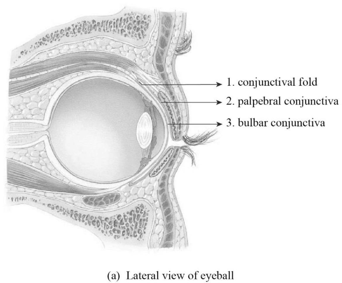
Fig1: The parts of conjunctiva
Explanation of Solution
1. Conjunctival fold: The conjunctiva is the mucous membrane. The eyelids’ inner surface and eyeballs’ exposed surface are lined by the conjunctiva. The pocket is formed by the conjunctival fold that possesses contacts from moving toward the eyeball’s posterior part.
2. Palpebral conjunctiva: The palpebral conjunctiva is the portion of the conjunctiva. The interior of the eyelid is covered by the palpebral conjunctiva.
3. Bulbar conjunctiva: The bulbar conjunctiva is otherwise referred to as ocular conjunctiva and it is a portion of the conjunctiva. The bulbar conjunctiva does not cover the cornea. It covers the anterior portion of the white of the eye.
To label: The structures of the lacrimal apparatus in Figure 24.1 (b).
Answer to Problem 1.1BGL
Pictorial representation:
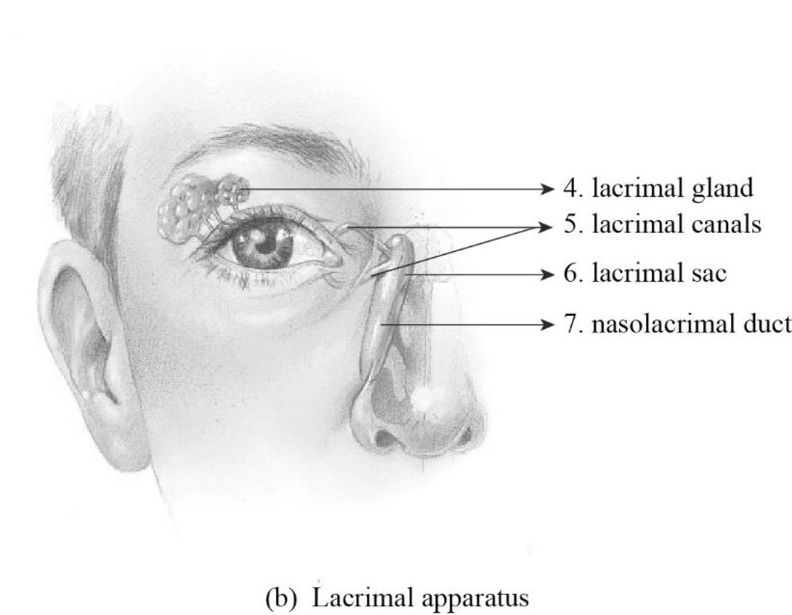
Fig1: The structures of the lacrimal apparatus
Explanation of Solution
A group of structure involved in draining and generating tears is referred to as the lacrimal apparatus.
4. Lacrimal gland: The lacrimal gland secretes and generates tears onto the surface of the eye. The fluid is released continuously by the lacrimal gland, which protects and cleanses the surface of the eyes.
5. Lacrimal canals: They are small channels found in every eyelid and it is also called as lacrimal canaliculi. The tear is drained into the enlarged lacrimal sac from the eyes by the lacrimal canals.
6. Lacrimal sac: The dilated oval upper end of the nasolacrimal duct is lacrimal sac. The lacrimal sac is also referred to as lachrymal sac.
7. Nasolacrimal duct: The nasolacrimal duct is otherwise known as the tear duct. The tear is received by the enlarged nasolacrimal duct from the lacrimal sac and the tears are drained into the nasal cavity.
Want to see more full solutions like this?
Chapter 24 Solutions
Laboratory Manual for Anatomy and Physiology, 6e Loose-Leaf Print Companion with WileyPLUS Blackboard Card Set


- An 1100 pound equine patient was given 20 mg/kg sucralfate 3 times a day, 2.8 mg/kg famotidine twice a day, and 10mg/kg doxycycline twice a day. Sucralfate comes as a 1 gm tablet, famotidine as 20 mg tablets, and doxycycline as 100mg tablets. All are in bottles of 100 tablets.How many total mg are needed for the patient and how many tablets of each would be needed to provide each dose?How many bottles of each would be needed to have available if this patient were to be on this drug regimen for 5 days?arrow_forwardThe patient needs a solution of 2.5% dextrose in Lactated Ringer’s solution to run at 75 ml/hr for at least the next 12hours. LRS comes in fluid bags of 500 ml, 1 Liter, 3 Liters and 5 Liters. How can a 2.5% solution be made by adding50% dextrose to the LRS?arrow_forward“Gretchen” was a 68-pound canine who came to the VMTH as small animal surgery patient. She receivedacepromazine, 0.2 mg/kg from a 10 mg/ml solution and oxymorphone, 0.08 mg/kg from a 1 mg/ml solution before surgery.What are the mechanisms of action of acepromazine and oxymorphone? Why would they be given together?How many mg provide each dose and how many ml of each of these solutions were given?arrow_forward
- After surgery, “Gretchen” was put on carprofen, 1 mg/pound bid (twice a day). The tablets come in 25, 75 and 100 mgsizes. Which size tablet would be appropriate?What is the mechanism of action of carprofen?An outpatient prescription was written for her so she would have enough for 10 days. How many tablets did she need?What information needs to be on her out-patient prescription?arrow_forwardJoden Koepp olor in chickens is due to incomplete dominance. BB = Black chicken, WW = White BLOOD TYPES Arhite chicken is In humans, Rh positive blood is dominant (R) over Rh negative blood (r). A man with type 0, Rh positive blood (whose mother had Rh negative blood), marries a woman with type AB, Rh negative blood. Several children were born. is? R R Genotypes Phenotypes RRR RR Rr Rr 4/16 RR R RR RK Rr Rr 4/16 rr 3/4 Rh posi 1/4 Rh negu 1/2 Rr rr rr rrrr 88 888 75 e genotype of the man? the genotype of the woman? The mother of the man had type AB blood.arrow_forwardPlease indentify the unknown organismarrow_forward
- 5G JA ATTC 3 3 CTIA A1G5 5 GAAT I I3 3 CTIA AA5 Fig. 5-3: The Eco restriction site (left) would be cleaved at the locations indicated by the arrows. However, a SNP in the position shown in gray (right) would prevent cleavage at this site by EcoRI One of the SNPs in B. rapa is found within the Park14 locus and can be detected by RFLP analysis. The CT polymorphism is found in the intron of the Bra013780 gene found on Chromosome 1. The Park14 allele with the "C" in the SNP has two EcoRI sites and thus is cleaved twice by EcoRI If there is a "T" in that SNP, one of the EcoRI sites is altered, so the Park14 allele with the T in the SNP has only one EcoRI site (Fig. 5-3). Park14 allele with SNP(C) Park14 allele with SNPT) 839 EcoRI 1101 EcoRI 839 EcoRI Fig. 5.4: Schematic restriction maps of the two different Park14 alleles (1316 bp long) of B. rapa. Where on these maps is the CT SNP located? 90 The primers used to amplify the DNA at the Park14 locus (see Fig. 5 and Table 3 of Slankster et…arrow_forwardFrom your previous experiment, you found that this enhancer activates stripe 2 of eve expression. When you sequence this enhancer you find several binding sites for the gap gene, Giant. To test how Giant interacts with eve, you decide to remove all of the Giant binding sites from the eve enhancer. What results do you expect to see with respect to eve expression?arrow_forwardWhat experiment could you do to see if the maternal gene, bicoid, is sufficient to form anterior fates?arrow_forward
 Human Anatomy & Physiology (11th Edition)BiologyISBN:9780134580999Author:Elaine N. Marieb, Katja N. HoehnPublisher:PEARSON
 Biology 2eBiologyISBN:9781947172517Author:Matthew Douglas, Jung Choi, Mary Ann ClarkPublisher:OpenStax
 Anatomy & PhysiologyBiologyISBN:9781259398629Author:McKinley, Michael P., O'loughlin, Valerie Dean, Bidle, Theresa StouterPublisher:Mcgraw Hill Education,
 Molecular Biology of the Cell (Sixth Edition)BiologyISBN:9780815344322Author:Bruce Alberts, Alexander D. Johnson, Julian Lewis, David Morgan, Martin Raff, Keith Roberts, Peter WalterPublisher:W. W. Norton & Company
 Laboratory Manual For Human Anatomy & PhysiologyBiologyISBN:9781260159363Author:Martin, Terry R., Prentice-craver, CynthiaPublisher:McGraw-Hill Publishing Co.
 Inquiry Into Life (16th Edition)BiologyISBN:9781260231700Author:Sylvia S. Mader, Michael WindelspechtPublisher:McGraw Hill Education