Concept explainers
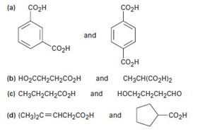
How would you use NMR (either 13C or 1H) to distinguish between the following pairs of isomers?

a)

Interpretation:
How NMR (either 13C or 1H) could be used to distinguish between benzene-1,2-dicarboxylic acid and benzene-1,3-dicarboxylic acid is to be stated.
Concept introduction:
In 1HNMR, the acidic COOH proton normally absorbs as singlet, broadened in few cases, near 12 δ.
In 13CNMR, the carboxyl carbon absorb in the range 165-185 δ. The aromatic and α, β-unsaturated acids absorb near 165 δ and saturated aliphatic acids near 185 δ. The nitrile carbons absorb in the range 115-130 δ.
Compounds with symmetrical structures (like p-disubstituted phenyl) will have a simpler spectrum.
To state:
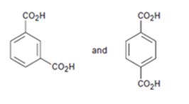
How NMR (either 13C or 1H) could be used to distinguish between benzene-1,3-dicarboxylic acid and benzene-1,4-dicarboxylic acid.
Answer to Problem 55AP
1,4-Benzenedicarboxylic acid will have a simpler spectrum. In 1H NMR, there will be a doublet of doublet in the aromatic region and a singlet for carboxyl proton around 12 δ. In 13C NMR there will be three signals, two in aromatic region and one for carboxyl carbon around 165 δ.
The 1H NMR of 1,3-Benzenedicarboxylic acid will have a multiplet in the aromatic region due to the ring protons and a singlet for carboxyl proton around 12 δ. In 13C NMR there will be five signals, four in aromatic region and one for carboxyl carbon around 165 δ.
Explanation of Solution
1,4-Benzenedicarboxylic acid has a symmetrical structure. Hence it gives minimum peaks in both 1H NMR and 13C NMR spectrum.
In 1H NMR, the four aromatic hydrogens belong to two groups and thus give a doublet of doublet in the aromatic region. The carboxyl proton gives another signal. Thus there will be three signals.
In 13C NMR, the six ring carbons classify themselves into two groups and give two signals while the carboxyl carbons give a signal. Thus there will be three signals.
1,3-Benzenedicarboxylic acid does not have a symmetrical structure. Hence it gives more peaks in both 1H NMR and 13C NMR spectrum than 1,4-Benzenedicarboxylic acid.
In 1H NMR, the four aromatic hydrogens give complex multiplet signals in the aromatic region. The carboxyl protons yield another signal. Thus there will be two signals.
In 13C NMR, the six ring carbons classify themselves into four groups and give four signals while the carboxyl carbons give a signal. Thus there will be five signals.
1,4-Benzenedicarboxylic acid will have a simpler spectrum. In 1H NMR, there will be a doublet of doublet in the aromatic region and a singlet for carboxyl proton around 12 δ. In 13C NMR there will be three signals, two in aromatic region and one for carboxyl carbon around 165 δ.
The 1H NMR of 1,3-Benzenedicarboxylic acid will have a multiplet in the aromatic region due to the ring protons and a singlet for carboxyl proton around 12 δ. In 13C NMR there will be five signals, four in aromatic region and one for carboxyl carbon around 165 δ.
b) HO2CCH2CH2CO2H and CH3CH(CO2H)2
Interpretation:
How NMR (either 13C or 1H) could be used to distinguish between succinic acid and ethane-1,1-dicarboxylic acid is to be stated.
Concept introduction:
In 1HNMR, the acidic COOH proton normally absorbs as singlet, broadened in few cases, near 12 δ.
In 13CNMR, the carboxyl carbon absorb in the range 165-185 δ. The aromatic and α, β-unsaturated acids absorb near 165 δ and saturated aliphatic acids near 185 δ. The nitrile carbons absorb in the range 115-130 δ.
Compounds with symmetrical structures (like p-disubstituted phenyl) will have a simpler spectrum.
To state:
How NMR (either 13C or 1H) could be used to distinguish between succinic acid and ethane-1,1-dicarboxylic acid.
Answer to Problem 55AP
Succinic acid will have a simpler spectrum. In 1H NMR, there will be two signals, a triplet for the methylene groups and a singlet for carboxyl protons around 12 δ. In 13C NMR there will be two signals, one for the methylene carbon and one for carboxyl carbons around 165 δ.
The 1H NMR, 2-methylmalonic acid will have three signals, a doublet for the methyl protons, a quartet for the methane proton and a singlet for carboxyl proton around 12 δ. In 13C NMR there will be three signals, one for methyl carbon another for the methine carbon and a third one for carboxyl carbons around 165 δ.
Explanation of Solution
Succinic acid has a simpler spectrum since it is symmetrical. In 1H NMR, the two methylene protons split each other giving a triplet integrating to four protons. The two carboxyl protons yield another singlet signal. Thus there will be two signals a triplet and a singlet.
In 13C NMR, the four carbons in succinic acid classify themselves into two groups and give two signals. Thus there will be two signals, one for methylene carbons and other for carboxyl carbons.
2-methylmalonicacid does not have a symmetrical structure. Hence it gives more peaks in both 1H NMR and 13C NMR spectrum than succinic acid.
In 1H NMR, there will be a doublet for the methyl protons, a quartet integrating to one proton for the methine proton signal and a singlet for the carboxyl protons. Thus there will be three signals.
In 13C NMR, there will be separate signals for methyl carbon, methine carbon and carboxyl carbons. Thus there will be three signals.
Succinic acid will have a simpler spectrum. In 1H NMR, there will be two signals, a triplet for the methylene groups and a singlet for carboxyl protons around 12 δ. In 13C NMR there will be two signals, one for the methylene carbons and one for carboxyl carbons around 165 δ.
The 1H NMR, 2-methylmalonic acid will have three signals, a doublet for the methyl protons, a quartet for the methine proton and a singlet for carboxyl proton around 12 δ. In 13C NMR there will be three signals, one for methyl carbon another for the methine carbon and a third one for carboxyl carbons around 165 δ.
c) CH3CH2CH2CO2H and HOCH2CH2CH2CHO
Interpretation:
How NMR (either 13C or 1H) could be used to distinguish between succinic acid and 4-hydroxybutanal is to be stated.
Concept introduction:
In 1HNMR, the acidic COOH proton normally absorbs as singlet, broadened in few cases, near 12 δ.
In 13CNMR, the carboxyl carbon absorb in the range 165-185 δ. The aromatic and α, β-unsaturated acids absorb near 165 δ and saturated aliphatic acids near 185 δ. The nitrile carbons absorb in the range 115-130 δ.
Compounds with symmetrical structures will have a simpler spectrum.
In 1HNMR the aldehyde protons absorb near 10 δ with a coupling constant, J=3Hz. Hydrogens on the carbon next to aldehyde group absorb near 2.0-2.3 δ.
In 13CNMR, saturated aldehydes and ketones usually absorb in the region from 200 to 215 δ while the aromatic and unsaturated carbonyl compounds absorb in the 190 to 200 δ region.
To state:
How NMR (either 13C or 1H) could be used to distinguish between butanoic acid and 4-hydroxybutanal.
Answer to Problem 55AP
In 1H NMR of butanoic acid there will be a signal around 12 δ while 1H NMR of 3-hydroxybutanal there will be a signal for the aldehydic proton around 9-10 δ and for the hydroxyl proton in the range 3.0-4.5 δ. There will be no signal around 12 δ. In 13C NMR the carboxyl carbon absorbs around 165 δ while the absorption due to aldehydic carbon is observed around 200-215 δ.
Explanation of Solution
The two compounds can be easily distinguished. For butanoic acid there will be signals for the absorption of carboxyl proton in 1H NMR (12 δ) and carboxyl carbon in 13C NMR (165 δ). In 3-hydroxybutanal, there will be signals for the absorption of hydroxyl proton in 1H NMR (3.0-4.5 δ) and aldehydic proton around 9-10 δ. The aldehydic carbon will resonate in 13C NMR in the range 200-215 δ.
In 1H NMR of butanoic acid there will be a signal around 12 δ while 1H NMR of 3-hydroxybutanal there will be a signal for the aldehydic proton around 9-10 δ and for the hydroxyl proton in the range 3.0-4.5 δ. There will be no signal around 12 δ. In 13C NMR the carboxyl carbon absorbs around 165 δ while the absorptrion due to aldehydic carbon is observed around 200-215 δ.
d)

Interpretation:
How NMR (either 13C or 1H) could be used to distinguish between 4-methylpent-3-eneoic acid and cyclopentylcarboxylic acid is to be stated.
Concept introduction:
In 1HNMR, the acidic COOH proton normally absorbs as singlet, broadened in few cases, near 12 δ.
In 13CNMR, the carboxyl carbon absorb in the range 165-185 δ. The aromatic and α, β-unsaturated acids absorb near 165 δ and saturated aliphatic acids near 185 δ. The nitrile carbons absorb in the range 115-130 δ.
Alkenes show a signal in the range 4.5-6.5 δ in 1HNMR and around 110-150 δ in 13CNMR.
Compounds with symmetrical structures will have a simpler spectrum.
To state:
How NMR (either 13C or 1H) could be used to distinguish between 4-methylpent-3-eneoic acid and cyclopentylcarboxylic acid.
Answer to Problem 55AP
In 1HNMR, 4-methylpent-3-eneoic acid will show the peak due to olefinic proton absorption in the range 4.5-6.5 δ. In 13CNMR, the two carbons in the double bond each will give a signal in the range 110-150 δ. There will be no signals in these ranges for cyclopentanecarboxylic acid in both the spectra.
Explanation of Solution
4-Methylpent-3-eneoic acid has a hydrogen atom attached to olefinic double bond. Since it is a vinylic hydrogen its absorption peak is observed in the range 4.5-6.5 δ. The two olefinic carbons are not equivalent. Hence they give two signals in the range 110-150 δ. In the 1HNMR and 13CNMR spectra of cyclopentanecarboxylic acid no signals will be observed in the ranges 4.5-6.5 δ and 110-150 δ respectively.
In 1HNMR, 4-methylpent-3-eneoic acid will show the peak due to olefinic proton absorption in the range 4.5-6.5 δ. In 13CNMR, the two carbons in the double bond each will give a signal in the range 110-150 δ. There will be no signals in these ranges for cyclopentanecarboxylic acid in both the spectra.
Want to see more full solutions like this?
Chapter 20 Solutions
Organic Chemistry - With Access (Custom)
- Using reaction free energy to predict equilibrium composition Consider the following equilibrium: 2NOCI (g) 2NO (g) + Cl2 (g) AGº =41. kJ Now suppose a reaction vessel is filled with 4.50 atm of nitrosyl chloride (NOCI) and 6.38 atm of chlorine (C12) at 212. °C. Answer the following questions about this system: ? rise Under these conditions, will the pressure of NOCI tend to rise or fall? x10 fall Is it possible to reverse this tendency by adding NO? In other words, if you said the pressure of NOCI will tend to rise, can that be changed to a tendency to fall by adding NO? Similarly, if you said the pressure of NOCI will tend to fall, can that be changed to a tendency to rise by adding NO? yes no If you said the tendency can be reversed in the second question, calculate the minimum pressure of NO needed to reverse it. Round your answer to 2 significant digits. 0.035 atm ✓ G 00. 18 Ararrow_forwardHighlight each glycosidic bond in the molecule below. Then answer the questions in the table under the drawing area. HO- HO- -0 OH OH HO NG HO- HO- OH OH OH OH NG OHarrow_forward€ + Suppose the molecule in the drawing area below were reacted with H₂ over a platinum catalyst. Edit the molecule to show what would happen to it. That is, turn it into the product of the reaction. Also, write the name of the product molecule under the drawing area. Name: ☐ H C=0 X H- OH HO- H HO- -H CH₂OH ×arrow_forward
- Draw the Haworth projection of the disaccharide made by joining D-glucose and D-mannose with a ẞ(1-4) glycosidic bond. If the disaccharide has more than one anomer, you can draw any of them. Click and drag to start drawing a structure. Xarrow_forwardEpoxides can be opened in aqueous acid or aqueous base to produce diols (molecules with two OH groups). In this question, you'll explore the mechanism of epoxide opening in aqueous acid. 2nd attempt Be sure to show all four bonds at stereocenters using hash and wedge lines. 0 0 Draw curved arrows to show how the epoxide reacts with hydronium ion. 100 +1: 1st attempt Feedback Be sure to show all four bonds at stereocenters using hash and wedge lines. See Periodic Table See Hint H A 5 F F Hr See Periodic Table See Hintarrow_forward03 Question (1 point) For the reaction below, draw both of the major organic products. Be sure to consider stereochemistry. > 1. CH₂CH₂MgBr 2. H₂O 3rd attempt Draw all four bonds at chiral centers. Draw all stereoisomers formed. Draw the structures here. e 130 AN H See Periodic Table See Hint P C Brarrow_forward
- You may wish to address the following issues in your response if they are pertinent to the reaction(s) you propose to employ:1) Chemoselectivity (why this functional group and not another?) 2) Regioselectivity (why here and not there?) 3) Stereoselectivity (why this stereoisomer?) 4) Changes in oxidation state. Please make it in detail and draw it out too in what step what happens. Thank you for helping me!arrow_forward1) Chemoselectivity (why this functional group and not another?) 2) Regioselectivity (why here and not there?) 3) Stereoselectivity (why this stereoisomer?) 4) Changes in oxidation state. Everything in detail and draw out and write it.arrow_forwardCalculating the pH at equivalence of a titration 3/5 Izabella A chemist titrates 120.0 mL of a 0.7191M dimethylamine ((CH3)2NH) solution with 0.5501 M HBr solution at 25 °C. Calculate the pH at equivalence. The pk of dimethylamine is 3.27. Round your answer to 2 decimal places. Note for advanced students: you may assume the total volume of the solution equals the initial volume plus the volume of HBr solution added. pH = ☐ ✓ 18 Ar Boarrow_forward
- Alcohols can be synthesized using an acid-catalyzed hydration of an alkene. An alkene is combined with aqueous acid (e.. sulfuric acid in water). The reaction mechanism typically involves a carbocation intermediate. > 3rd attempt 3343 10 8 Draw arrows to show the reaction between the alkene and hydronium ion. that 2nd attempt Feedback 1st attempt تعمال Ju See Periodic Table See Hint F D Ju See Periodic Table See Hintarrow_forwardDraw the simplified curved arrow mechanism for the reaction of acetone and CHgLi to give the major product. 4th attempt Π Draw the simplified curved arrow mechanism T 3rd attempt Feedback Ju See Periodic Table See Hint H -H H -I H F See Periodic Table See Hintarrow_forwardSelect the correct reagent to accomplish the first step of this reaction. Then draw a mechanism on the Grignard reagent using curved arrow notation to show how it is converted to the final product. 4th attempt Part 1 (0.5 point) Select the correct reagent to accomplish the first step of this reaction. Choose one: OA Mg in ethanol (EtOH) OB. 2 Li in THF O C. Li in THF D. Mg in THF O E Mg in H2O Part 2 (0.5 point) Br Part 1 Bri Mg CH B CH, 1 Draw intermediate here, but no arrows. © TE See Periodic Table See Hint See Hint ין Harrow_forward

 Introduction to General, Organic and BiochemistryChemistryISBN:9781285869759Author:Frederick A. Bettelheim, William H. Brown, Mary K. Campbell, Shawn O. Farrell, Omar TorresPublisher:Cengage Learning

