
Videos
The five figures to the right are labeled a through e. Fill in the blanks with the proper identification.
(a) —————-
(b)—————-
(d)—————-
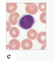
(e)—————-
To review:
Identification of the given cell structures in the five figures.
Introduction:
The WBC (white blood cells) are the cells that provide defense from several pathogens and fight against foreign substances. These are of two types namely, granulocytes and agranulocytes. Granulocytes are basophils, eosinophils, and neutrophils. Agranulocytes are lymphocytes and monocytes.
Explanation of Solution
The structures of different cells are labeled as below:
Basophil
Lymphocyte
Neutrophils
Eosinophil
Monocyte
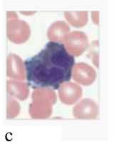
Basophils are the cells that assist the mast cells in the inflammation reaction. The cell is round and the nucleus is generally not visible due to the presence of large, dense purple granules in the cytoplasm. In the diagram also, the large dense purple–blue granules are clearly visible, which makes the cytoplasm difficult to be observed. It comprises heparin, which is an anticoagulant, prevents blood from clotting rapidly. Vasodilator histamine is also present in these cells, which are responsible for blood flow to tissues.
Lymphocytes are the cells of the lymphatic system that provides defense against specific toxins and pathogens. These cells have a large nucleus as well as the cytoplasm is very little. The cells present in the lymphatic system provides defense against certain toxins or pathogens.
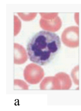
Neutrophils are the cells that are phagocytic in nature and engulf cellular debris. The cell is round and nucleus resembles a series of beads. Large pale inclusions are present in the cytoplasm. These cells are round and nucleus usually resembles the series of beads. The cytoplasm present is usually large and consists of pale inclusions.
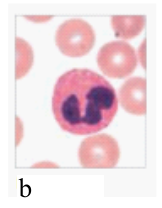
Eosinophil is a large cell with two lobes usually. Large granules are present in the cytoplasm. These granules stain orange–red with acidic dyes. The prime function is to attack anything, which carries antibody. It mostly plays an important role in fighting parasitic infections and leads to the suppression of inflammation.
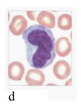
Monocytes are very large cells having a kidney-bean-shaped nucleus. Abundant pale cytoplasm is present in the cell. It enters damaged tissue and produces histamine along with other chemicals.
Therefore, the structures of the different cells can be named as
| a | Neutrophils |
| b | Eosinophil |
| c | Basophil |
| d | Monocyte |
| e | Lymphocyte |
Want to see more full solutions like this?
Chapter 20 Solutions
Human Anatomy (8th Edition) - Standalone book


- Select all of the following that the ablation (knockout) or ectopoic expression (gain of function) of Hox can contribute to. Another set of wings in the fruit fly, duplication of fingernails, ectopic ears in mice, excess feathers in duck/quail chimeras, and homeosis of segment 2 to jaw in Hox2a mutantsarrow_forwardSelect all of the following that changes in the MC1R gene can lead to: Changes in spots/stripes in lizards, changes in coat coloration in mice, ectopic ear formation in Siberian hamsters, and red hair in humansarrow_forwardPleiotropic genes are genes that (blank) Cause a swapping of organs/structures, are the result of duplicated sets of chromosomes, never produce protein products, and have more than one purpose/functionarrow_forward
- A loss of function mutation in Pitx1 enhancers can cause (blank) Removal of Pitx1 exons and growth of ectopic hindlimbs, growth of extra ectopic forelimbs, loss of forelimb specification and development, and loss of hindlimb specification and developmentarrow_forwardHox1a most likely contributes to (blank) patterning in the developing embryo? Ventral, posterior, limb or anteriorarrow_forwardSelect all of the following that can help establish Hox gene expression boundaries (things that affect Hox and not things that Hox affects). Retinoic acid, anterior/posterior axis, fibroblast growth factors, vagal neural crest, and enhancersarrow_forward
- Ectopic expression of Hox often results in (blank) phenotypes. (Blank) transformations are characterized by the replacement of one body part/structure with another. Hoxeotic, homealoneotic, joexotic, or homeoticarrow_forwardWhat's the difference when drawing omega-6 and omega-3?arrow_forward. Consider a base substitution mutation that occurred in a DNA sequence that resulted in a change in the encoded protein from the amino acid glutamic acid to aspartic acid. Normally the glutamic acid amino acid is located on the outside of the soluble protein but not near an active site. O-H¨ A. What type of mutation occurred? O-H B. What 2 types of chemical bonds are found in the R-groups of each amino acid? The R groups are shaded. CH2 CH2 CH2 H2N-C-COOH H2N-C-COOH 1 H Glutamic acid H Aspartic acid C. What 2 types of bonds could each R-group of each of these amino acids form with other molecules? D. Consider the chemical properties of the two amino acids and the location of the amino acid in the protein. Explain what effect this mutation will have on this protein's function and why.arrow_forward
- engineered constructs that consist of hollow fibers are acting as synthetic capillaries, around which cells have been loaded. The cellular space around a single fiber can be modeled as if it were a Krogh tissue cylinder. Each fiber has an outside “capillary” radius of 100 µm and the “tissue” radius can be taken as 200 µm. The following values apply to the device:R0 = 20 µM/secaO2 = 1.35 µM/mmHgDO2,T = 1.67 x 10⁻⁵ cm2/secPO2,m = 4 x 10⁻³ cm/secInstead of blood inside the fibers, the oxygen transport and tissue consumption are being investigated by usingan aqueous solution saturated with pure oxygen. As a result, there is no mass transfer resistance in the synthetic“capillary”, only that due to the membrane itself. Rather than accounting for pO2 variations along the length ofthe fiber, use an average value in the “capillary” of 130 mmHg.Is the tissue fully oxygenated?arrow_forwardMolecular Biology Please help with question. thank you You are studying the expression of the lac operon. You have isolated mutants as described below. In the presence of glucose, explain/describe what would happen, for each mutant, to the expression of the lac operon when you add lactose AND what would happen when the bacteria has used up all of the lactose (if the mutant is able to use lactose).5. Mutations in the lac operator that strengthen the binding of the lac repressor 200 fold 6. Mutations in the promoter that prevent binding of RNA polymerase 7. Mutations in CRP/CAP protein that prevent binding of cAMP8. Mutations in sigma factor that prevent binding of sigma to core RNA polymerasearrow_forwardMolecular Biology Please help and there is an attached image. Thank you. A bacteria has a gene whose protein/enzyme product is involved with the synthesis of a lipid necessary for the synthesis of the cell membrane. Expression of this gene requires the binding of a protein (called ACT) to a control sequence (called INC) next to the promoter. A. Is the expression/regulation of this gene an example of induction or repression?Please explain:B. Is this expression/regulation an example of positive or negative control?C. When the lipid is supplied in the media, the expression of the enzyme is turned off.Describe one likely mechanism for how this “turn off” is accomplished.arrow_forward

- Surgical Tech For Surgical Tech Pos CareHealth & NutritionISBN:9781337648868Author:AssociationPublisher:Cengage
 Principles Of Radiographic Imaging: An Art And A ...Health & NutritionISBN:9781337711067Author:Richard R. Carlton, Arlene M. Adler, Vesna BalacPublisher:Cengage Learning
 Human Physiology: From Cells to Systems (MindTap ...BiologyISBN:9781285866932Author:Lauralee SherwoodPublisher:Cengage Learning