
Videos
To label: The structures given in Fig 18.1
Concept introduction: Neurons are the fundamental functioning units of the nervous system. Neurons share many similar features with other body cells; only one different feature is that they can conduct signals throughout the body.
Answer to Problem 1.1BGL
Pictorial representation:
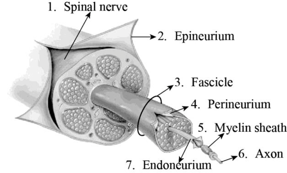
Fig 1: Transverse section displaying the coverings of a spinal nerve
Explanation of Solution
The spinal nerves function like “telephone lines” and bring back and forth messages between the body and the spinal cord to regulate motions as well as sensations. There are two roots in each spinal nerve; they are the ventral root and the dorsal root. An axon is a lengthy, slender projection that emerges from a nerve cell. The myelin sheath is formed of a fatty material that surrounds the nerve cells.
The dense and uneven connective tissue that surrounds the peripheral nerves is termed as the epineurium. This tissue envelops several blood vessels that supply blood to the nerves and nerve fascicles. The epineurium is abundantly seen in the regions of joints. Hence, they protect the nerves from injury as well as stretching.
A nerve fascicle is otherwise known as fasciculus. This structure is formed of a bundle of axons. These fascicles are present in the peripheral nervous system. They are bundled together with the blood vessels and fatty tissues.
Nerve fascicles are enveloped by a kind of connective tissue that is termed as the perineurium. The perineurium is formed of perineurial cells, and they are also referred to as myoepithelioid in some cases. The perineurium membrane is a smooth, tube-like transparent structure that could be easily detached from the fibers which it surrounds.
The connective tissue that surrounds the unmyelinated and myelinated axons is called the endoneurium. The endoneurium is otherwise known as the endoneurial channel or Henle’s sheath. It is delicate and surrounds the myelin sheath of every myelinated nerve fiber. The endoneurium is made up of endoneurial cells.
To label: The structures given in Fig 18.2
Answer to Problem 1.1BGL
Pictorial representation:
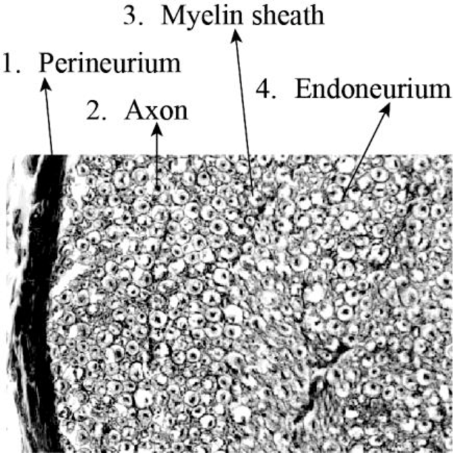
Fig 2: A transverse section photomicrograph through a spinal nerve fascicle
Explanation of Solution
An axon is a long, slim projection that emerges from a neuron. The myelin sac consists of a fat material that surrounds the cells of the nerves. The endoneurium is also known as the Henle’s sheath or endoneurial channel. It is fragile and surrounds every myelinated nerve fiber’s sheath. The endoneurium consists of endoneurial cells.
Want to see more full solutions like this?
Chapter 18 Solutions
Laboratory Manual for Anatomy and Physiology, 6e Loose-Leaf Print Companion


- Ch.21 What causes patients infected with the yellow fever virus to turn yellow (jaundice)? A. low blood pressure and anemia B. excess leukocytes C. alteration of skin pigments D. liver damage in final stage of disease — What is the advantage for malarial parasites to grow and replicate in red blood cells? A. able to spread quickly B. able to avoid immune detection C. low oxygen environment for growth D. cooler area of the body for growth — Which microbe does not live part of its lifecycle outside humans? A. Toxoplasma gondii B. Cytomegalovirus C. Francisella tularensis D. Plasmodium falciparum — explain your answer thoroughlyarrow_forwardCh.22 Streptococcus pneumoniae has a capsule to protect it from killing by alveolar macrophages, which kill bacteria by… A. cytokines B. antibodies C. complement D. phagocytosis — What fact about the influenza virus allows the dramatic antigenic shift that generates novel strains? A. very large size B. enveloped C. segmented genome D. over 100 genes — explain your answer thoroughlyarrow_forwardWhat is this?arrow_forward
- Molecular Biology A-C components of the question are corresponding to attached image labeled 1. D component of the question is corresponding to attached image labeled 2. For a eukaryotic mRNA, the sequences is as follows where AUGrepresents the start codon, the yellow is the Kozak sequence and (XXX) just represents any codonfor an amino acid (no stop codons here). G-cap and polyA tail are not shown A. How long is the peptide produced?B. What is the function (a sentence) of the UAA highlighted in blue?C. If the sequence highlighted in blue were changed from UAA to UAG, how would that affecttranslation? D. (1) The sequence highlighted in yellow above is moved to a new position indicated below. Howwould that affect translation? (2) How long would be the protein produced from this new mRNA? Thank youarrow_forwardMolecular Biology Question Explain why the cell doesn’t need 61 tRNAs (one for each codon). Please help. Thank youarrow_forwardMolecular Biology You discover a disease causing mutation (indicated by the arrow) that alters splicing of its mRNA. This mutation (a base substitution in the splicing sequence) eliminates a 3’ splice site resulting in the inclusion of the second intron (I2) in the final mRNA. We are going to pretend that this intron is short having only 15 nucleotides (most introns are much longer so this is just to make things simple) with the following sequence shown below in bold. The ( ) indicate the reading frames in the exons; the included intron 2 sequences are in bold. A. Would you expected this change to be harmful? ExplainB. If you were to do gene therapy to fix this problem, briefly explain what type of gene therapy youwould use to correct this. Please help. Thank youarrow_forward
- Molecular Biology Question Please help. Thank you Explain what is meant by the term “defective virus.” Explain how a defective virus is able to replicate.arrow_forwardMolecular Biology Explain why changing the codon GGG to GGA should not be harmful. Please help . Thank youarrow_forwardStage Percent Time in Hours Interphase .60 14.4 Prophase .20 4.8 Metaphase .10 2.4 Anaphase .06 1.44 Telophase .03 .72 Cytukinesis .01 .24 Can you summarize the results in the chart and explain which phases are faster and why the slower ones are slow?arrow_forward
- Can you circle a cell in the different stages of mitosis? 1.prophase 2.metaphase 3.anaphase 4.telophase 5.cytokinesisarrow_forwardWhich microbe does not live part of its lifecycle outside humans? A. Toxoplasma gondii B. Cytomegalovirus C. Francisella tularensis D. Plasmodium falciparum explain your answer thoroughly.arrow_forwardSelect all of the following that the ablation (knockout) or ectopoic expression (gain of function) of Hox can contribute to. Another set of wings in the fruit fly, duplication of fingernails, ectopic ears in mice, excess feathers in duck/quail chimeras, and homeosis of segment 2 to jaw in Hox2a mutantsarrow_forward
 Human Anatomy & Physiology (11th Edition)BiologyISBN:9780134580999Author:Elaine N. Marieb, Katja N. HoehnPublisher:PEARSON
 Biology 2eBiologyISBN:9781947172517Author:Matthew Douglas, Jung Choi, Mary Ann ClarkPublisher:OpenStax
 Anatomy & PhysiologyBiologyISBN:9781259398629Author:McKinley, Michael P., O'loughlin, Valerie Dean, Bidle, Theresa StouterPublisher:Mcgraw Hill Education,
 Molecular Biology of the Cell (Sixth Edition)BiologyISBN:9780815344322Author:Bruce Alberts, Alexander D. Johnson, Julian Lewis, David Morgan, Martin Raff, Keith Roberts, Peter WalterPublisher:W. W. Norton & Company
 Laboratory Manual For Human Anatomy & PhysiologyBiologyISBN:9781260159363Author:Martin, Terry R., Prentice-craver, CynthiaPublisher:McGraw-Hill Publishing Co.
 Inquiry Into Life (16th Edition)BiologyISBN:9781260231700Author:Sylvia S. Mader, Michael WindelspechtPublisher:McGraw Hill Education