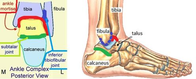
Watch this video (http://openstaxcollege.org/l/anklejoint1) for a tutorial on the anatomy of the anklejoint. What are the three ligaments found on the lateral sideof the ankle joint?
Histology
Histology is the microanatomy method and a branch of biology that studies the anatomy of tissues. It includes viewing tissue in a magnified view under the microscope. Microanatomy also includes the process of study of organs called organology and the study of cells called cytology. Histopathology is a branch of biology that includes microscopic identification of diseased tissue. The field of histology comprises the preparation of the tissues and collection of cells as specimens for examination under the microscope. These processes are done by technicians like histologists, histotechnicians, and biomedical scientists. Histopathology is the diagnosis and research of tissue diseases that require the examination of tissues and/or cells under a microscope. Histopathologists are in charge of determining tissue diagnosis and assisting clinicians in managing a patient's care.
Endocrine System
Human body functions due to the collective work of the organ systems. One of them is the endocrine system. It is a chemical messenger system constituting the hormones directly released by the endocrine glands into the circulatory system. The study of this system is known as endocrinology. The word 'endon' means inside, and 'crine' means secrete, making the word "endocrine."
Watch this video (http://openstaxcollege.org/l/
anklejoint1) for a tutorial on the anatomy of the ankle
joint. What are the three ligaments found on the lateral side
of the ankle joint?

Three bones participate in the formation of the ankle- Tibia, fibula and Talus. The malleoli, medial malleolus(bony process extanding distaly off the medial tibia)and lateral malleolus(distal-most aspect of the fibula) , along with their supporting ligament stabilize the talus underneath the tibia.The ankle is composed of three joints-the talocrural joint( Ankle mortise), the subtalar joint and the Inferior tibiofibular joint.
Step by step
Solved in 2 steps with 2 images


