describe the functional anatomy of the ureters, urinarybladder, and male and female urethra; and
describe the functional anatomy of the ureters, urinary
bladder, and male and female urethra; and
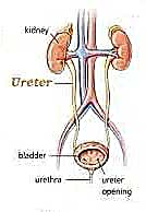
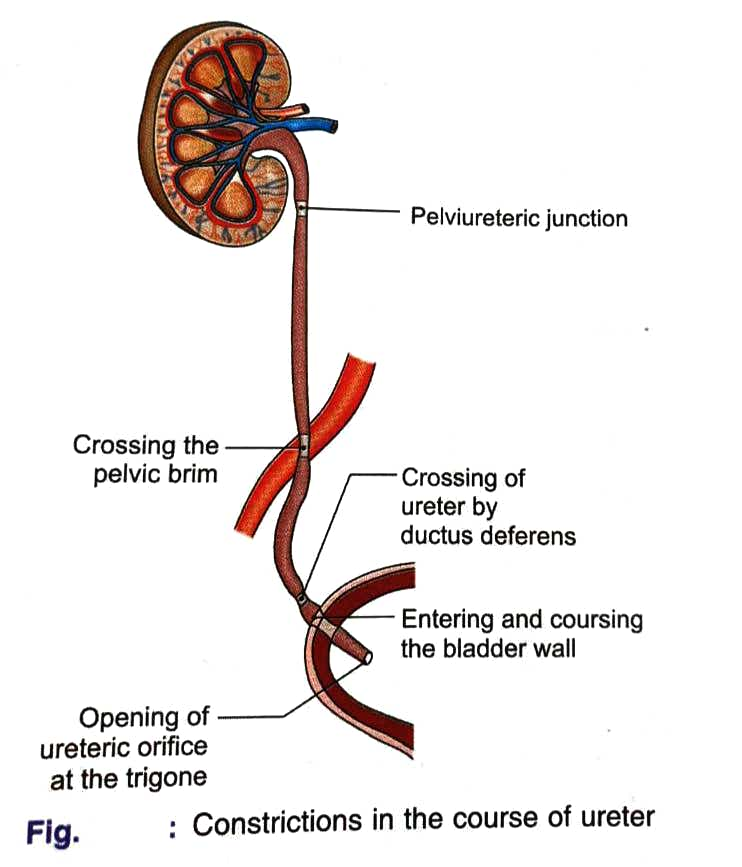
FUNCTIONAL ANATOMYNOF URETERS : the ureters are a pair of narrow , thick walled muscular tubes which convey urine from the kidneys to the urinary bladder . They lie deep to the peritoneum closely applied to the posterior abdominal wall in the upper part , and to the lateral pelvic wall in the lower part . The intravesicle oblique course of the ureter has a valvular action and prevents regurgitation of urine from the bladder to the ureter .

Ureter is composed of the innermost mucous membrane . middle layer of well developed smooth muscle layer and outer tunica adventitia .

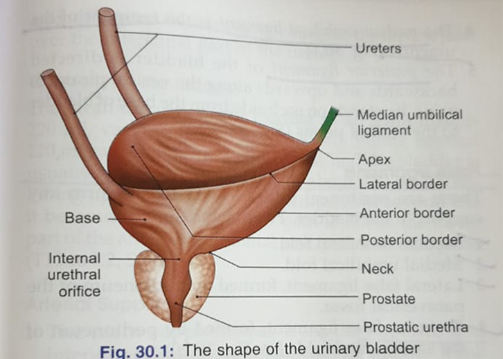
FUNCTIONAL ANATOMY OF URINARY BLADDER : urinary bladder is a muscular reservoir of urine , which lies in the anterior part of the pelvic cavity . It varies in size , shape and position according to the amount of urine it contains . When empty it lies entirely within the pelvis; but as it fills it expands and extends upwards into the abdominal cavity , reaching up to the umbilicus or even higher .
diagram showing urinary bladder

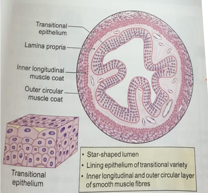
The epithelium of urinary bladder is of transitional variety . The luminal cells are well defined dome shaped squamous cells with prominent nuclei . The middle layers are pear shaped cells and the basal layer is of short columnar cells . The inner muscle coat is admixture of longitudinal , circular and oblique layers . Outermost layer is the serous or adventitial coat .
diagram showing histology of urinary bladder

Step by step
Solved in 2 steps with 7 images


