3. Tyrosine kinase receptors are pairs of proteins that span the plasma membrane. On the extracellular side of the membrane, one or more sites are present that bind to signaling ligands such as insulin or growth factors. On the intracellular side, the end of peptide chains on each protein phosphorylate the other member of the pair, providing active docking sites that initiate cellular responses. The signal is switched off by dissociation of the ligand. For each ligand-receptor system, the equilibrium constant, k, controls the distribution of receptor-bound and unbound ligands. In systems with large values of k, a site is likely to be occupied, even at low concentrations of ligand. When k is small, the likelihood of binding is low, even when the concentration of ligand is high. To initiate a new stimulus response cycle for the receptor, the ligand must dissociate. Larger values of k mean that the receptor is more likely to be occupied and thus unavailable to bind another ligand. Some ligand- binding systems have multiple binding sites. For example, hemoglobin binds four oxygen molecules, whereas myoglobin has only a single binding site. When multiple binding sites are present, the presence of an already-bound ligand can cooperatively affect the binding of other ligands on the same protein. For hemoglobin, the binding is positively cooperative. The affinity of oxygen for heme increases as the number of bound oxygen molecules increases. Part A: Describe the features in this graph for hemoglobin that demonstrate positive cooperativity. Number of sites bound Concentration of ligand Hemoglobin (positive cooperativity) k = 10 (negative cooperativity) Myoglobin k = 1 (negative cooperativity) k = 0.01 (negative cooperativity) Part B: The insulin receptor (IR) is a tyrosine kinase receptor that has two sites to which insulin can attach. IR is negatively cooperative. In the graph in (A), the dependence of the bound fraction on available insulin is similar to the curve for with negative cooperativity Describe the features of this oure in the graph that demonstrate negative cooperativity
3. Tyrosine kinase receptors are pairs of proteins that span the plasma membrane. On the extracellular side of the membrane, one or more sites are present that bind to signaling ligands such as insulin or growth factors. On the intracellular side, the end of peptide chains on each protein phosphorylate the other member of the pair, providing active docking sites that initiate cellular responses. The signal is switched off by dissociation of the ligand. For each ligand-receptor system, the equilibrium constant, k, controls the distribution of receptor-bound and unbound ligands. In systems with large values of k, a site is likely to be occupied, even at low concentrations of ligand. When k is small, the likelihood of binding is low, even when the concentration of ligand is high. To initiate a new stimulus response cycle for the receptor, the ligand must dissociate. Larger values of k mean that the receptor is more likely to be occupied and thus unavailable to bind another ligand. Some ligand- binding systems have multiple binding sites. For example, hemoglobin binds four oxygen molecules, whereas myoglobin has only a single binding site. When multiple binding sites are present, the presence of an already-bound ligand can cooperatively affect the binding of other ligands on the same protein. For hemoglobin, the binding is positively cooperative. The affinity of oxygen for heme increases as the number of bound oxygen molecules increases. Part A: Describe the features in this graph for hemoglobin that demonstrate positive cooperativity. Number of sites bound Concentration of ligand Hemoglobin (positive cooperativity) k = 10 (negative cooperativity) Myoglobin k = 1 (negative cooperativity) k = 0.01 (negative cooperativity) Part B: The insulin receptor (IR) is a tyrosine kinase receptor that has two sites to which insulin can attach. IR is negatively cooperative. In the graph in (A), the dependence of the bound fraction on available insulin is similar to the curve for with negative cooperativity Describe the features of this oure in the graph that demonstrate negative cooperativity
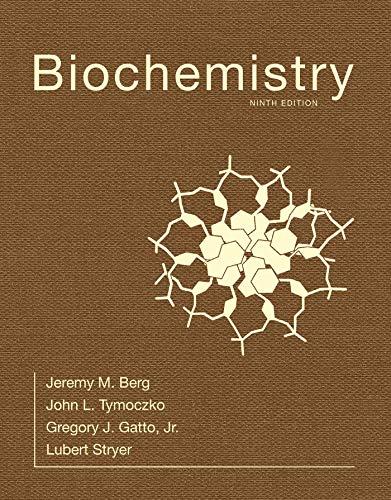
Biochemistry
9th Edition
ISBN:9781319114671
Author:Lubert Stryer, Jeremy M. Berg, John L. Tymoczko, Gregory J. Gatto Jr.
Publisher:Lubert Stryer, Jeremy M. Berg, John L. Tymoczko, Gregory J. Gatto Jr.
Chapter1: Biochemistry: An Evolving Science
Section: Chapter Questions
Problem 1P
Related questions
Question
Part B

Transcribed Image Text:3. Tyrosine kinase receptors are pairs of proteins that span the plasma membrane. On the extracellular side of the membrane, one or more sites are present that bind to signaling ligands such as insulin or growth factors. On the intracellular side, the ends
of peptide chains on each protein phosphorylate the other member of the pair, providing active docking sites that initiate cellular responses. The signal is switched off by dissociation of the ligand. For each ligand-receptor system, the equilibrium
constant, k, controls the distribution of receptor-bound and unbound ligands. In systems with large values of k, a site is likely to be occupied, even at low concentrations of ligand. When k is small, the likelihood of binding is low, even when the
concentration of ligand is high. To initiate a new stimulus response cycle for the receptor, the ligand must dissociate. Larger values of k mean that the receptor is more likely to be occupied and thus unavailable to bind another ligand. Some ligand-
binding systems have multiple binding sites. For example, hemoglobin binds four oxygen molecules, whereas myoglobin has only a single binding site. When multiple binding sites are present, the presence of an already-bound ligand can cooperatively
affect the binding of other ligands on the same protein. For hemoglobin, the binding is positively cooperative. The affinity of oxygen for heme increases as the number of bound oxygen molecules increases.
Part A: Describe the features in this graph for hemoglobin that demonstrate positive cooperativity.
Number of sites bound
0
Concentration of ligand
Hemoglobin (positive cooperativity)
k = 10 (negative cooperativity)
Myoglobin
k = 1 (negative cooperativity)
k = 0.01 (negative cooperativity)
Part B: The insulin receptor (IR) is a tyrosine kinase receptor that has two sites to which insulin can attach. IR is negatively cooperative. In the graph in (A), the dependence of the bound fraction on available insulin is similar to the curve for with
negative cooperativity. Describe the features of this curve in the graph that demonstrate negative cooperativity.
Expert Solution
This question has been solved!
Explore an expertly crafted, step-by-step solution for a thorough understanding of key concepts.
Step by step
Solved in 4 steps

Recommended textbooks for you

Biochemistry
Biochemistry
ISBN:
9781319114671
Author:
Lubert Stryer, Jeremy M. Berg, John L. Tymoczko, Gregory J. Gatto Jr.
Publisher:
W. H. Freeman

Lehninger Principles of Biochemistry
Biochemistry
ISBN:
9781464126116
Author:
David L. Nelson, Michael M. Cox
Publisher:
W. H. Freeman

Fundamentals of Biochemistry: Life at the Molecul…
Biochemistry
ISBN:
9781118918401
Author:
Donald Voet, Judith G. Voet, Charlotte W. Pratt
Publisher:
WILEY

Biochemistry
Biochemistry
ISBN:
9781319114671
Author:
Lubert Stryer, Jeremy M. Berg, John L. Tymoczko, Gregory J. Gatto Jr.
Publisher:
W. H. Freeman

Lehninger Principles of Biochemistry
Biochemistry
ISBN:
9781464126116
Author:
David L. Nelson, Michael M. Cox
Publisher:
W. H. Freeman

Fundamentals of Biochemistry: Life at the Molecul…
Biochemistry
ISBN:
9781118918401
Author:
Donald Voet, Judith G. Voet, Charlotte W. Pratt
Publisher:
WILEY

Biochemistry
Biochemistry
ISBN:
9781305961135
Author:
Mary K. Campbell, Shawn O. Farrell, Owen M. McDougal
Publisher:
Cengage Learning

Biochemistry
Biochemistry
ISBN:
9781305577206
Author:
Reginald H. Garrett, Charles M. Grisham
Publisher:
Cengage Learning

Fundamentals of General, Organic, and Biological …
Biochemistry
ISBN:
9780134015187
Author:
John E. McMurry, David S. Ballantine, Carl A. Hoeger, Virginia E. Peterson
Publisher:
PEARSON